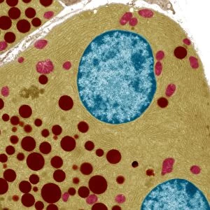
Metal Print : Pancreas cell, TEM


Resolution may be poor at this size (110 DPI)
![]()
Metal Prints from Science Photo Library
Pancreas cell, TEM
Pancreas cell. Coloured transmission electron micrograph (TEM) of an acinar (exocrine) pancreatic cell. Acinar cells secrete the inactive precursors (zymogens) of digestive enzymes to the small intestine, via the pancreatic ducts. The enzymes are secreted in vesicles known as zymogen granules (not seen) and are activated using proteolysis in the small intestine to aid the breakdown of carbohydrates, fats and proteins. The production and secretion of the zymogens are controlled by the cells endoplasmic reticulum and golgi apparatus (blue/red). The cells nucleus (purple, centre) and mitochondria (yellow) are visible
Science Photo Library features Science and Medical images including photos and illustrations
Media ID 6450923
© STEVE GSCHMEISSNER/SCIENCE PHOTO LIBRARY
Acinar Cell Body Cytological Cytology Digestive Endoplasmic Reticulum Enzymatic Enzyme Enzymes Exocrine False Colour Golgi Apparatus Inactive Magnified Image Micrograph Microscopic Photos Nucleus Pancreas Pancreatic Precursor Secretory Storage Subjects Transmission Electron Microgra Transmission Electron Microscope Transport Zymogen False Coloured
20"x24" (61x51cm) Metal Print
Discover the intricacy of life with Media Storehouse's Metal Prints featuring this stunning Transmission Electron Micrograph (TEM) image of a Pancreas cell from Science Photo Library. Witness the complexity of an acinar (exocrine) pancreatic cell, which plays a crucial role in the secretion of digestive enzymes. Our high-quality Metal Prints bring the microscopic world to life with vibrant colors and exceptional detail, making for a captivating addition to any scientific or medical space. Order yours today and bring the beauty of science into your home or office.
Made with durable metal and luxurious printing techniques, our metal photo prints go beyond traditional canvases, adding a cool, modern touch to your space. Wall mount on back. Eco-friendly 100% post-consumer recycled ChromaLuxe aluminum surface. The thickness of the print is 0.045". Featuring a Scratch-resistant surface and Rounded corners. Backing hangers are attached to the back of the print and float the print 1/2-inch off the wall when hung, the choice of hanger may vary depending on size and International orders will come with Float Mount hangers only. Finished with a brilliant white high gloss surface for unsurpassed detail and vibrance. Printed using Dye-Sublimation and for best care we recommend a non-ammonia glass cleaner, water, or isopropyl (rubbing) alcohol to prevent harming the print surface. We recommend using a clean, lint-free cloth to wipe off the print. The ultra-hard surface is scratch-resistant, waterproof and weatherproof. Avoid direct sunlight exposure.
Made with durable metal and luxurious printing techniques, metal prints bring images to life and add a modern touch to any space
Estimated Image Size (if not cropped) is 60.9cm x 50.8cm (24" x 20")
Estimated Product Size is 61.5cm x 51.4cm (24.2" x 20.2")
These are individually made so all sizes are approximate
Artwork printed orientated as per the preview above, with landscape (horizontal) orientation to match the source image.
EDITORS COMMENTS
This stunning coloured transmission electron micrograph (TEM) captures the intricate beauty of a pancreas cell. Specifically, it showcases an acinar pancreatic cell, which plays a crucial role in our digestive system. These cells secrete inactive precursors called zymogens that eventually transform into digestive enzymes and aid in breaking down carbohydrates, fats, and proteins. The image reveals the inner workings of the cell with remarkable detail. The zymogen granules responsible for enzyme secretion are not visible but are known to be present within these cells. The endoplasmic reticulum and golgi apparatus, depicted in blue and red respectively, control the production and release of these zymogens. In this magnified view, we can also observe the nucleus at the center of the cell represented in purple while mitochondria appear as yellow structures. This false-coloured representation allows us to appreciate both their distinct features and their vital roles within cellular function. Overall, this photograph provides a glimpse into the microscopic world of cytology by showcasing one component of our complex biological system – highlighting its normal anatomy and healthy state. It serves as a reminder of how intricately designed our bodies are on even the smallest scale.
MADE IN THE USA
Safe Shipping with 30 Day Money Back Guarantee
FREE PERSONALISATION*
We are proud to offer a range of customisation features including Personalised Captions, Color Filters and Picture Zoom Tools
SECURE PAYMENTS
We happily accept a wide range of payment options so you can pay for the things you need in the way that is most convenient for you
* Options may vary by product and licensing agreement. Zoomed Pictures can be adjusted in the Cart.