Fraud Blocker Skip to main content

Enzymatic Collection

"Unlocking the Secrets of Enzymatic Power: Exploring Ricin A-chain and Hammerhead Ribozyme Molecules" Enzymes

Background imageEnzymatic Collection: Ricin A-chain, artwork C017 / 3653

Ricin A-chain, artwork C017 / 3653
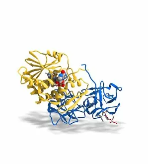
Ricin A-chain. Computer artwork showing the enzymatically active A-chain from a molecule of the toxic protein ricin. Ricin comprises two entwined amino acid chains; A (seen here) and B (not shown)

Background imageEnzymatic Collection: Ricin molecule, artwork C017 / 3652

Ricin molecule, artwork C017 / 3652
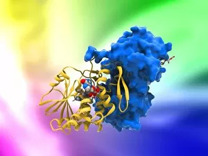
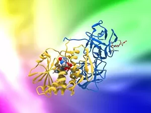
Ricin molecule. Computer artwork showing the structure of a molecule of the toxic protein ricin. Ricin comprises two entwined amino acid chains; A (yellow) and B (blue)

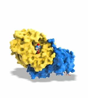
Background imageEnzymatic Collection: Ricin molecule, artwork C017 / 3651

Ricin molecule, artwork C017 / 3651
Ricin molecule. Computer artwork showing the structure of a molecule of the toxic protein ricin. Ricin comprises two entwined amino acid chains; A (yellow) and B (blue)

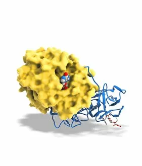
Background imageEnzymatic Collection: Ricin molecule, artwork C017 / 3650

Ricin molecule, artwork C017 / 3650
Ricin molecule. Computer artwork showing the structure of a molecule of the toxic protein ricin. Ricin comprises two entwined amino acid chains; A (yellow) and B (blue)

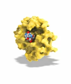
Background imageEnzymatic Collection: Ricin A-chain, artwork C017 / 3654

Ricin A-chain, artwork C017 / 3654
Ricin A-chain. Computer artwork showing the enzymatically active A-chain from a molecule of the toxic protein ricin. Ricin comprises two entwined amino acid chains; A (seen here) and B (not shown)

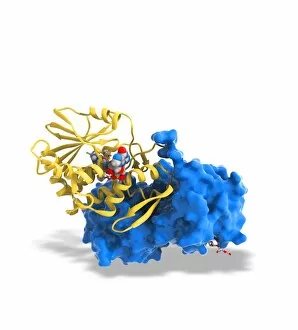
Background imageEnzymatic Collection: Ricin molecule, artwork C017 / 3649

Ricin molecule, artwork C017 / 3649
Ricin molecule. Computer artwork showing the structure of a molecule of the toxic protein ricin. Ricin comprises two entwined amino acid chains; A (yellow) and B (blue)

Background imageEnzymatic Collection: Exosome complex, molecular model

Exosome complex, molecular model. This multi-protein complex functions to break up strands of RNA (ribonucleic acid, pink) during biochemical processes

Background imageEnzymatic Collection: Ricin molecule, artwork C017 / 3656

Ricin molecule, artwork C017 / 3656
Ricin molecule. Computer artwork showing the structure of a molecule of the toxic protein ricin. Ricin comprises two entwined amino acid chains; A (yellow) and B (blue)

Background imageEnzymatic Collection: Ricin molecule, artwork C017 / 3655

Ricin molecule, artwork C017 / 3655
Ricin molecule. Computer artwork showing the structure of a molecule of the toxic protein ricin. Ricin comprises two entwined amino acid chains; A (yellow) and B (blue)

Background imageEnzymatic Collection: Ricin molecule, artwork C017 / 3648

Ricin molecule, artwork C017 / 3648
Ricin molecule Computer artwork showing the structure of a molecule of the toxic protein ricin (blue and yellow) with an active ribosome in the background

Background imageEnzymatic Collection: Heterotrimeric G protein complex C013 / 7186

Heterotrimeric G protein complex C013 / 7186
Heterotrimeric G protein complex, molecular model showing secondary structure. Also called the large G proteins, these activate enzymes and metabolic pathways

Background imageEnzymatic Collection: Pancreas cell, TEM

Pancreas cell, TEM
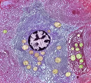
Pancreas cell. Coloured transmission electron micrograph (TEM) of an acinar (exocrine) pancreatic cell. Acinar cells secrete the inactive precursors (zymogens)

Background imageEnzymatic Collection: Secretory cells in pancreas, SEM

Secretory cells in pancreas, SEM
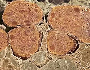
Pancreatic secretory cells. Coloured scanning electron micrograph (SEM) of a freeze-fracture through a healthy pancreas, showing the secretory tissue

Background imageEnzymatic Collection: Pancreas cells, SEM

Pancreas cells, SEM
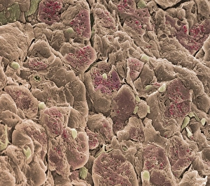
Pancreas cells. Coloured scanning electron micrograph (SEM) of acinar (exocrine) pancreatic cells. Acinar cells produce and excrete digestive enzymes to the small intestine, via the pancreatic ducts

Background imageEnzymatic Collection: DNA and restriction enzyme, artwork

DNA and restriction enzyme, artwork
DNA and restriction enzyme. Computer artwork of double-stranded DNA (deoxyribonucleic acid, blue) and a restriction enzyme protein EcoKI (green)

Background imageEnzymatic Collection: Pancreas cell, SEM

Pancreas cell, SEM
Pancreas cell. Coloured scanning electron micrograph (SEM) of an acinar (exocrine) pancreatic cell. Acinar cells produce and excrete digestive enzymes to the small intestine

Background imageEnzymatic Collection: Protein translation, artwork

Protein translation, artwork
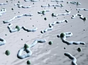
Protein translation. Artwork showing the process of translation, the final stage of the production of proteins from the genetic code

Background imageEnzymatic Collection: Cholinesterase enzyme

Cholinesterase enzyme. Molecular model of the secondary structure of butyrylcholinesterase (BChE), showing alpha helices (blue) and beta sheets (red and yellow)

Background imageEnzymatic Collection: Hammerhead ribozyme molecule

Hammerhead ribozyme molecule
Hammerhead ribozyme, molecular model. Ribozymes are RNA (ribonucleic acid) molecules that catalyse certain biochemical reactions

Background imageEnzymatic Collection: ATPase muscle enzyme

ATPase muscle enzyme
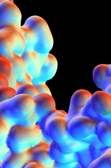
Calcium pumping ATPase enzyme. Computer model of an electrostatic potential surface map of part of the ATPase enzyme that pumps calcium in and out of muscle cells and controls muscle contractions


All Professionally Made to Order for Quick Shipping

"Unlocking the Secrets of Enzymatic Power: Exploring Ricin A-chain and Hammerhead Ribozyme Molecules" Enzymes, the tiny molecular machines that drive countless biological processes, continue to fascinate scientists. Among them, the enigmatic Ricin A-chain and Hammerhead ribozyme molecules stand out as captivating subjects of study. In artwork C017 / 3653, we delve into the intricate structure of Ricin A-chain. This potent toxin derived from castor beans possesses enzymatic activity that can disrupt protein synthesis within cells. Its complex architecture holds clues to its deadly mechanism. Artwork C017 / 3652 showcases another perspective on the Ricin molecule's composition. With its distinctive shape and arrangement of atoms, it exemplifies nature's remarkable ability to create powerful enzymes with specific functions. Moving forward in our exploration, artwork C017 / 3649 presents an intriguing view of a ricin molecule at a different angle. Each representation offers fresh insights into this enzyme's potential applications in various fields such as medicine or biotechnology. Meanwhile, exosome complexes take center stage in our investigation with their vital roles in cell-to-cell communication and waste disposal systems. These intricate molecular models are depicted here as fascinating networks of proteins and RNA strands (artwork not specified). Returning to ricin-related studies, artworks C017 / 3656 and C017 / 3655 provide further glimpses into this enzyme's structural intricacies. As researchers unravel its secrets piece by piece, they inch closer towards harnessing its power for beneficial purposes while ensuring safety precautions. Amidst all these discoveries lies artwork C017 / 3648—a depiction showcasing yet another facet of ricin's complexity—highlighting how each new finding adds depth to our understanding. Lastly but equally compelling is the Hammerhead ribozyme molecule—an RNA-based catalyst capable of cleaving other RNA molecules like a pair of molecular scissors.