
Secretory Collection
"Unveiling the Hidden Secrets: Exploring the Intricacies Cells and Glands" Delving deep into the intricate workings of our body


All Professionally Made to Order for Quick Shipping
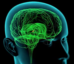
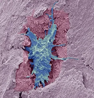
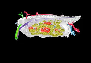
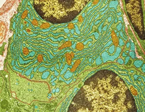
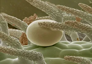
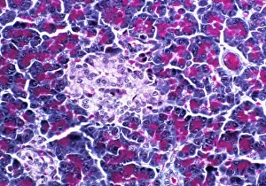
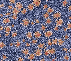
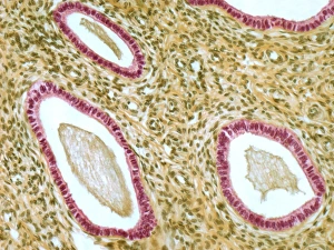
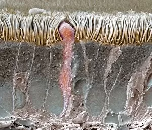
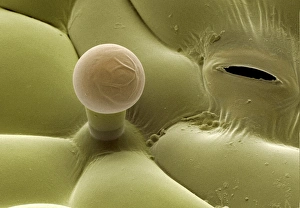
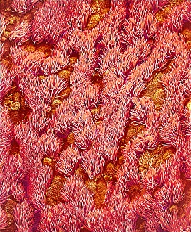
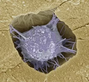
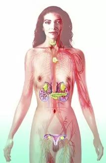
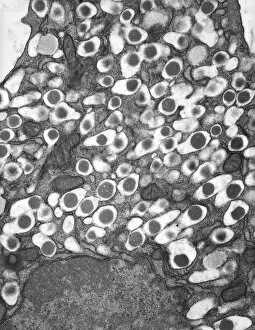
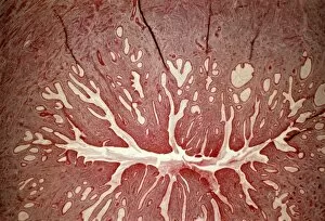
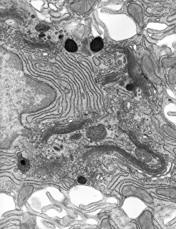
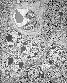
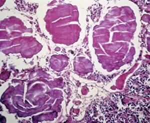
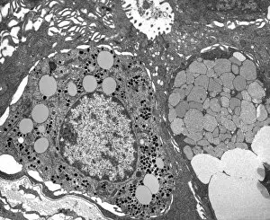
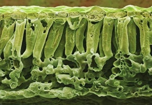
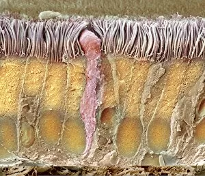
"Unveiling the Hidden Secrets: Exploring the Intricacies Cells and Glands" Delving deep into the intricate workings of our body, we uncover a world hints that hold immense significance. From the medulla oblongata in the brain to osteocyte bone cells, each structure plays a vital role in maintaining our well-being. Intriguing artwork showcases the complexity of these secretory systems. The medulla oblongata, often referred to as the "brain's control center, " orchestrates various bodily functions with its network of neurons. Meanwhile, osteocyte bone cells reveal their delicate intricacy under SEM C016 / 9025, highlighting their crucial role in bone formation and maintenance. Moving on to another remarkable organ, we encounter an artistic representation of pancreas anatomy. This gland houses pancreatic islets of Langerhans - clusters of specialized cells responsible for secreting hormones like insulin and glucagon that regulate blood sugar levels. Venturing further into microscopic realms, TEM unveils plasma cells - guardians against infections by producing antibodies that defend our immune system. Thyme leaf oil glands capture attention with their unique secretion properties while contributing to aromatic experiences. SEM images offer glimpses into hidden worlds within us - brain lining displaying its intricate web-like structure; fallopian tube cells showcasing their elegant arrangement; nasal lining revealing its finely textured surface; cervix exhibiting distinctive features under light micrograph F006 / 9805; trachea lining demonstrating its protective capabilities through SEM C013 / 7126. These captivating visualizations remind us of the awe-inspiring complexity present within our bodies' secretory systems. They serve as a testament to nature's ingenuity and inspire us to delve deeper into understanding these hidden secrets that contribute so profoundly to our overall health and well-being.