Fraud Blocker Skip to main content

Enzymes Collection

Background imageEnzymes Collection: Glutamine synthetase enzyme

Glutamine synthetase enzyme computer model. This is a ligase enzyme, which forms chemical bonds between molecules. The different colours show the different subunits that comprise the protein

Background imageEnzymes Collection: Eosinophil white blood cell, TEM

Eosinophil white blood cell, TEM
Eosinophil white blood cell. Transmission electron micrograph (TEM) of a section through an eosinophil white blood cell (leukocyte)

Background imageEnzymes Collection: Pancreatic acinar cell

Pancreatic acinar cell

Background imageEnzymes Collection: X-ray crystallography C016 / 3824

X-ray crystallography C016 / 3824
X-ray crystallography. Researcher using an X-ray machine to obtain crystal diffraction patterns of proteins for 3-D imaging of enzymes

Background imageEnzymes Collection: Flap endonuclease protein F007 / 9914

Flap endonuclease protein F007 / 9914
Molecular model of the flap endonuclease protein

Background imageEnzymes Collection: Flap endonuclease protein F007 / 9916

Flap endonuclease protein F007 / 9916
Molecular model of the flap endonuclease protein

Background imageEnzymes Collection: Pancreatic exocrine cells, TEM

Pancreatic exocrine cells, TEM
Pancreatic exocrine cells

Background imageEnzymes Collection: Pancreatic acinar cell, TEM

Pancreatic acinar cell, TEM
Pancreatic acinar cell

Background imageEnzymes Collection: Drug effect on viruses, conceptual image C016 / 6253

Drug effect on viruses, conceptual image C016 / 6253
Drug effect on viruses, conceptual image

Background imageEnzymes Collection: Islet of Langerhans, light micrograph C016 / 0514

Islet of Langerhans, light micrograph C016 / 0514
Islet of Langerhans. Light micrograph of a section through an islet of Langerhans in a pancreas

Background imageEnzymes Collection: Islet of Langerhans, light micrograph C016 / 0513

Islet of Langerhans, light micrograph C016 / 0513
Islet of Langerhans. Light micrograph of a section through an islet of Langerhans in a pancreas

Background imageEnzymes Collection: Stilbene synthase molecule C014 / 2292

Stilbene synthase molecule C014 / 2292
Stilbene synthase, molecular model. This enzyme is produced by some plants (including grapes, peanuts and blueberries) in response to stress from either ultraviolet light or certain fungi

Background imageEnzymes Collection: Stilbene synthase molecule C014 / 2291

Stilbene synthase molecule C014 / 2291
Stilbene synthase, molecular model. This enzyme is produced by some plants (including grapes, peanuts and blueberries) in response to stress from either ultraviolet light or certain fungi

Background imageEnzymes Collection: Saliva chemicals, molecular model

Saliva chemicals, molecular model
Saliva chemicals. Molecular structure of a group of saliva molecules. These include the antibody immunoglobulin A (blue, double-y shape, see C014/5652)

Background imageEnzymes Collection: Biofuel production and use, diagram

Biofuel production and use, diagram. At far left the first stage is the source material (biomass, plant or other organic material). This is added to a separation tank (centre left)

Background imageEnzymes Collection: Alexander Fleming, caricature

Alexander Fleming, caricature
Alexander Fleming (1881-1955). Caricature of the Scottish biologist and pharmacologist Alexander Fleming holding a Petri dish

Background imageEnzymes Collection: Kidney tissue, fluorescence micrograph C016 / 8484

Kidney tissue, fluorescence micrograph C016 / 8484
Kidney tissue. Fluorescence deconvolution micrograph of a section through a kidney, showing glomeruli (green), cell nuclei (blue dots), and renal tubules (red, circular)

Background imageEnzymes Collection: X-ray crystallography C016 / 3823

X-ray crystallography C016 / 3823
X-ray crystallography. Researcher using an X-ray machine to obtain crystal diffraction patterns of proteins for 3-D imaging of enzymes

Background imageEnzymes Collection: DNA and methyltransferase complex C014 / 0011

DNA and methyltransferase complex C014 / 0011
DNA and methyltransferase complex. Molecular model showing a molecule of methyltransferase bound to a DNA (deoxyribonucleic acid) strand (red and yellow)

Background imageEnzymes Collection: HRas enzyme molecule C014 / 0010

HRas enzyme molecule C014 / 0010
HRas enzyme molecule. Molecular model showing the structure of a molecule of the enzyme GTPase HRas, also known as transforming protein p21

Background imageEnzymes Collection: NADH dehydrogenase molecule, artwork C014 / 0009

NADH dehydrogenase molecule, artwork C014 / 0009
NADH dehydrogenase molecule. Computer artwork showing the structure of a molecule of NADH dehydrogenase (NADH:ubiquinone reductase or Complex I)

Background imageEnzymes Collection: DNA replication by helicase enzyme C013 / 9382

DNA replication by helicase enzyme C013 / 9382
Computer artwork of DNA Helicase breaking apart the hydrogen bonds of a DNA strand for replication. Helicases are a class of enzymes vital to all living organisms

Background imageEnzymes Collection: DNA polymerase molecule C013 / 7909

DNA polymerase molecule C013 / 7909
DNA polymerase. Molecular model of a molecule of DNA polymerase (blue) replicating a strand of DNA (deoxyribonucleic acid, pink and turquoise). The secondary structure of the DNA polymerase is shown

Background imageEnzymes Collection: Venus flytrap digestive glands, SEM

Venus flytrap digestive glands, SEM
Venus flytrap digestive glands. Coloured scanning electron micrograph (SEM) of digestive glands from a Venus flytrap plant (Dionaea muscipula)

Background imageEnzymes Collection: Metopus protozoan

Metopus protozoan

Background imageEnzymes Collection: Thalassomyxa australis protozoan

Thalassomyxa australis protozoan
Thalassomyxa protozoan. Coloured scanning electron micrograph (SEM) of a Thalassomyxa australis prot- ozoan (single-celled animal)

Background imageEnzymes Collection: Lembadion protozoan

Lembadion protozoan. Coloured scanning electron micrograph (SEM) of a Lembadion bullinum protozoan (single-celled animal)

Background imageEnzymes Collection: Tetrahymena protozoa

Tetrahymena protozoa. Immunofluorescent light micrograph of two Tetrahymena thermophila protozoa (single-celled animals). Nuclei are green, cell walls red and cilia (hairs) blue. T

Background imageEnzymes Collection: Dendrocometes protozoan

Dendrocometes protozoan. Coloured scanning electron micrograph (SEM) of a Dendrocometes paradoxus protozoan (single-celled animal)

Background imageEnzymes Collection: Pancreas cell, TEM

Pancreas cell, TEM
Pancreas cell. Coloured transmission electron micrograph (TEM) of an acinar (exocrine) pancreatic cell

Background imageEnzymes Collection: Secretory cells in pancreas, SEM

Secretory cells in pancreas, SEM
Pancreatic secretory cells. Coloured scanning electron micrograph (SEM) of a freeze-fracture through a healthy pancreas, showing the secretory tissue

Background imageEnzymes Collection: Pancreas cells, SEM

Pancreas cells, SEM
Pancreas cells. Coloured scanning electron micrograph (SEM) of acinar (exocrine) pancreatic cells. Acinar cells produce and excrete digestive enzymes to the small intestine, via the pancreatic ducts

Background imageEnzymes Collection: Salivary gland, light micrograph

Salivary gland, light micrograph
Salivary gland. Coloured light micrograph of a section through a sublingual salivary gland, which is situated directly under the tongue

Background imageEnzymes Collection: Pancreas cell, SEM

Pancreas cell, SEM
Pancreas cell. Coloured scanning electron micrograph (SEM) of an acinar (exocrine) pancreatic cell

Background imageEnzymes Collection: Viral RNA replication cycle, artwork

Viral RNA replication cycle, artwork
Viral RNA replication cycle

Background imageEnzymes Collection: Pancreas tissue, SEM

Pancreas tissue, SEM
Pancreas tissue. Coloured scanning electron micrograph (SEM) of fractured pancreas tissue, showing numerous acinar cells, containing secretory zymogen granules

Background imageEnzymes Collection: Protein translation, artwork

Protein translation, artwork
Protein translation. Artwork showing the process of translation, the final stage of the production of proteins from the genetic code

Background imageEnzymes Collection: DNA replication fork, artwork

DNA replication fork, artwork
DNA replication fork. Diagram showing the cyclic sequence (right) for replication of DNA (deoxyribonucleic acid). Details of the DNA are at left

Background imageEnzymes Collection: DNA replication process, diagram

DNA replication process, diagram
DNA replication process. Diagram showing various stages in the replication of DNA (deoxyribonucleic acid). The process starts (top) when initiator proteins (blue-grey) separate the strands of DNA

Background imageEnzymes Collection: Lysyl oxidase enzyme molecule

Lysyl oxidase enzyme molecule. Computer artwork showing the secondary structure of the enzyme lysyl oxidase (LOX)

Background imageEnzymes Collection: Kinase molecule, secondary structure

Kinase molecule, secondary structure
Kinase molecule, computer model

Background imageEnzymes Collection: Co-enzyme NAD (nicotinamide) crystals

Co-enzyme NAD (nicotinamide) crystals
Polarised light micrograph of crystals of the co- enzyme nicotinamide adenine dinucleotide (NAD). This is an extremely important and widespread co- enzyme found in nature

Background imageEnzymes Collection: Caspase 3 molecule

Caspase 3 molecule
Caspase-3 molecule. Computer artwork showing the secondary structure of a molecule of caspase-3

Background imageEnzymes Collection: Art of cytochrome oxidase enzyme

Art of cytochrome oxidase enzyme
Cytochrome oxidase enzyme. Illustration of a model of the enzyme cytochrome oxidase, important in respiration

Background imageEnzymes Collection: DNA polymerase, molecular model

DNA polymerase, molecular model
DNA polymerase. Computer model showing the structure of a DNA polymerase molecule (green)

Background imageEnzymes Collection: Elaidic acid, computer model

Elaidic acid, computer model
Elaidic acid. Computer model of a molecule of elaidic acid, a trans fatty acid. Atoms are represented as spheres and are colour-coded: carbon (green), hydrogen (white) and oxygen (red)

Background imageEnzymes Collection: Lysozome protein crystals

Lysozome protein crystals
Lysozyme enzyme crystals. Polarised light micro- graph of crystals of the enzyme lysozyme from the egg white (albumen) of a domestic chicken

Background imageEnzymes Collection: Linoleic acid, computer model

Linoleic acid, computer model
Linoleic acid. Computer model of a molecule of linoleic acid, an omega-6 essential fatty acid. Atoms are represented as spheres and are colour- coded; carbon (blue), hydrogen (gold) and oxygen (red)

Background imageEnzymes Collection: Caspase 1 molecule

Caspase 1 molecule
Caspase-1 molecule. Computer artwork showing the secondary structure of a molecule of caspase-1. Caspase-1 is a protease, an enzyme that cleaves proteins

Background imageEnzymes Collection: Fragment of a kinase molecule, artwork

Fragment of a kinase molecule, artwork
Fragment of a kinase molecule, computer model

Background imageEnzymes Collection: Trypsin molecule, computer artwork

Trypsin molecule, computer artwork
Trypsin molecule. Computer model of a molecule of the digestive enzyme trypsin

Background imageEnzymes Collection: Pyruvate dehydrogenase enzyme

Pyruvate dehydrogenase enzyme. Illustration of the molecular structure of the enzyme pyruvate de- hydrogenase, important in glycolysis

Background imageEnzymes Collection: Lipase molecule, secondary structure

Lipase molecule, secondary structure
Lipase molecule, computer model. Lipase is an enzyme that breaks lipids (fats) into fatty acids and glycerol

Background imageEnzymes Collection: Ribozyme molecule

Ribozyme molecule
Ribozyme. Computer model of a ribozyme molecule. Ribozymes are RNA (ribonucleic acid) molecules that catalyse certain biochemical reactions

Background imageEnzymes Collection: Art of structure of enzyme cytochrome C

Art of structure of enzyme cytochrome C
Cytochrome C enzyme. Illustration of a model of the enzyme cytochrome C, important in the process of respiration. Cytochromes are proteins often regarded as enzymes

Background imageEnzymes Collection: Cholinesterase enzyme

Cholinesterase enzyme. Molecular model of the secondary structure of butyrylcholinesterase (BChE), showing alpha helices (blue) and beta sheets (red and yellow)

Background imageEnzymes Collection: Ferroxidase enzyme, molecular model

Ferroxidase enzyme, molecular model
Ferroxidase enzyme. Molecular model showing two views of the secondary structure of the human enzyme ferroxidase, also known as ceruloplasmin. Copper atoms are represented as red spheres

Background imageEnzymes Collection: Cytidine deaminase, molecular model

Cytidine deaminase, molecular model
Cytidine deaminase. Computer model of the enzyme, activation-induced (cytidine) deaminase (AID)

Background imageEnzymes Collection: Oleic acid, computer model

Oleic acid, computer model
Oleic acid. Computer model of a molecule of oleic acid, a monounsaturated fatty acid. Atoms are represented as spheres and are colour-coded: carbon (green), hydrogen (white) and oxygen (red)

Background imageEnzymes Collection: Lysozyme molecule, computer artwork

Lysozyme molecule, computer artwork
Lysozyme molecule. Computer model of a molecule of the antibacterial enzyme lysozyme. Lysozyme is a widely distributed protein in human bodily fluids, including tears and saliva

Background imageEnzymes Collection: Alcohol dehydrogenase, molecular model

Alcohol dehydrogenase, molecular model. Alcohol dehydrogenase (ADH) is an enzyme that facilitates the break-down of alcohols in the body, which could otherwise be toxic

Background imageEnzymes Collection: Kinase molecule, computer artwork

Kinase molecule, computer artwork
Kinase molecule, computer model

Background imageEnzymes Collection: Cyclin-depenent kinase 5 molecule

Cyclin-depenent kinase 5 molecule
Cyclin-dependent kinase 5. Molecular model showing the secondary structure of the enzyme cylcin- dependent kinase 5 (cdk5)

Background imageEnzymes Collection: Gamma-linolenic acid, computer model

Gamma-linolenic acid, computer model
Gamma-linolenic acid. Computer model of a molecule of gamma-linolenic acid (GLA), an omega-6 essential fatty acid

Background imageEnzymes Collection: Pancreas acinus, SEM

Pancreas acinus, SEM
Pancreas acinus. Coloured scanning electron micrograph (SEM) of a freeze-fracture through an acinus (yellow) in the pancreas. An acinus is a collection of glandular epithelial cells

Background imageEnzymes Collection: FK506-binding protein molecule

FK506-binding protein molecule

Background imageEnzymes Collection: Lipase molecule

Lipase molecule. Computer model showing the secondary structure of lipase. Alpha-helices are blue and beta-sheets are purple

Background imageEnzymes Collection: DNA polymerase Klenow fragment

DNA polymerase Klenow fragment
Klenow fragment of DNA polymerase I

Background imageEnzymes Collection: Protein tyrosine phosphatase molecule

Protein tyrosine phosphatase molecule. Computer model of the secondary structure of an intermediate form of protein tyrosine phosphatase. Beta-sheets are purple and alpha-helices are blue

Background imageEnzymes Collection: Cyclin-dependent kinase 2 enzyme

Cyclin-dependent kinase 2 enzyme, molecular model. This enzyme is found in cells, where it is involved in regulating the cell cycle, the cycle of cell division and cell growth

Background imageEnzymes Collection: ATPase muscle enzyme

ATPase muscle enzyme
Calcium pumping ATPase enzyme. Computer model of an electrostatic potential surface map of part of the ATPase enzyme that pumps calcium in and out of muscle cells and controls muscle contractions

Background imageEnzymes Collection: Pepsin molecule

Pepsin molecule
Pepsin enzyme. Computer graphic of the protein- digesting enzyme pepsin. It is a protease enzyme that is secreted as part of gastric juice into the stomach in an inactive form known as pepsinogen


All products are expertly crafted, using premium materials, tailored to your specifications and promptly shipped


EDITORS COMMENTS

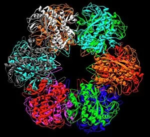
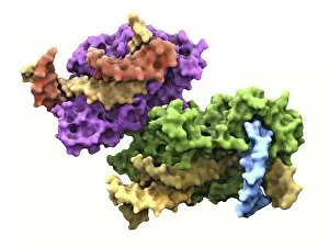
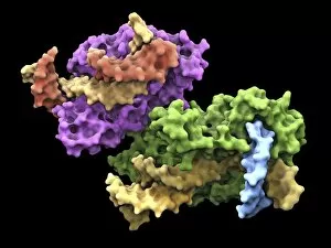
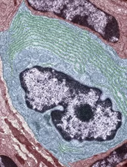
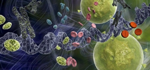
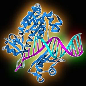
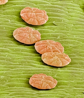
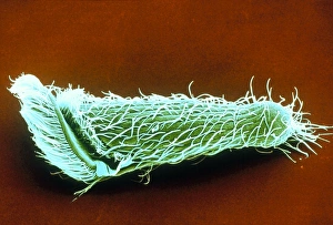
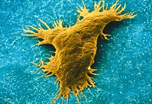
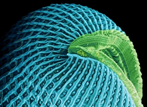
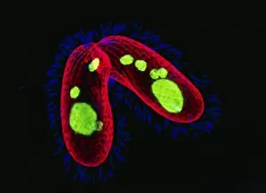
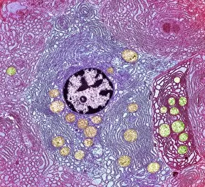
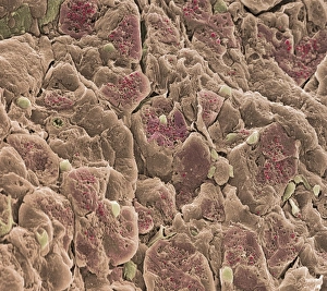
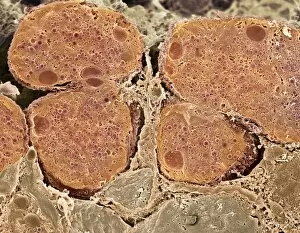
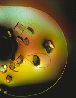
Enzymes: The Tiny Powerhouses of Life From the intricate Glutamine synthetase enzyme to the mesmerizing Lysozome protein crystals, enzymes never cease to amaze us. With X-ray crystallography C016 / 3824 revealing their hidden structures, we uncover a world of complexity and beauty. The Flap endonuclease proteins F007/9914 and F007/9916 play crucial roles in DNA repair, ensuring our genetic material remains intact. Meanwhile, the Ubiquitin activating enzyme protein E1 F007/9908 orchestrates cellular processes by tagging proteins for degradation or modification. Underneath the microscope's lens, Pancreatic exocrine cells reveal their secretory nature while Eosinophil white blood cells stand as guardians against foreign invaders. In this microscopic realm, Pancreatic acinar cells shine with their ability to produce digestive enzymes essential for nutrient breakdown. Through TEM images, we witness the bustling activity within these pancreatic acinar cells as they tirelessly fulfill their vital functions. And let's not forget about those resilient Eosinophil white blood cells that defend our bodies from harm and can truly remarkable entities that drive countless biochemical reactions necessary for life itself. Their diverse structures and functions make them indispensable players in maintaining homeostasis and sustaining living organisms. So next time you marvel at an enzymatic reaction or delve into the fascinating world of molecular biology, remember that behind every biological process lies an army working diligently to keep everything running smoothly.

© Copyright Media Storehouse, All Rights Reserved 2003 - 2025
Facebook     Pinterest     100% Payment Secure     Reviews IO