Photographic Print > Popular Themes > Human Body
Photographic Print : Nerve and glial cells, light micrograph

![]()
Photo Prints From Science Photo Library
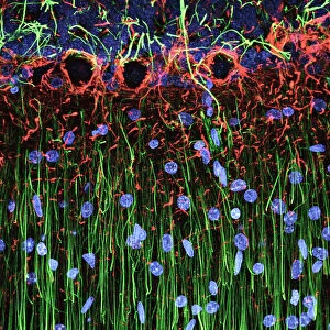
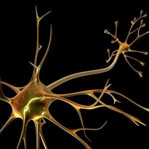
Nerve and glial cells, light micrograph
Nerve and glial cells, fluorescence light micrograph. These are neural stem cells that have differentiated into neurons (nerve cells, blue) and glial cells (support cells, red). The branching processes from the neurons are called dendrites. Fluorescent markers have been used to highlight proteins. The proteins stained here are beta III-tubulin (blue), a cytoskeleton element found in neurons, and GFAP (glial fibrillary acidic protein, red), forming the cytoskeleton of the glial cells. This sample is from rat tissue
Science Photo Library features Science and Medical images including photos and illustrations
Media ID 10948201
© DANIEL SCHROEN, CELL APPLICATIONS INC/SCIENCE PHOTO LIBRARY
Animal Body Astrocyte Astrocytes Cell Biology Cellular Cytoskeletal Cytoskeleton Fluorescence Fluorescence Micrograph Fluorescing Gfap Glial Cell Glial Fibrillary Acidic Protein Nerve Nerve Cell Neuron Neurone Neurones Neurons Nobody Proteins Stains Tubulin Brain Cells Light Micrograph Light Microscope Nervous System Neurological Neurology Protein
10"x8" Photo Print
Discover the intricacies of the human body with Media Storehouse's range of Photographic Prints. Feast your eyes on this stunning light micrograph image of Nerve and Glial Cells by Daniel Schroen, captured through the lens of Cell Applications Inc/Science Photo Library. Witness the beauty of neural stem cells as they transform into neurons, depicted in a captivating blue hue, and glial cells, shown in vibrant red, providing essential support. Invite the wonders of science into your home or office with this exquisite piece of scientific art.
Photo prints are produced on Kodak professional photo paper resulting in timeless and breath-taking prints which are also ideal for framing. The colors produced are rich and vivid, with accurate blacks and pristine whites, resulting in prints that are truly timeless and magnificent. Whether you're looking to display your prints in your home, office, or gallery, our range of photographic prints are sure to impress. Dimensions refers to the size of the paper in inches.
Our Photo Prints are in a large range of sizes and are printed on Archival Quality Paper for excellent colour reproduction and longevity. They are ideal for framing (our Framed Prints use these) at a reasonable cost. Alternatives include cheaper Poster Prints and higher quality Fine Art Paper, the choice of which is largely dependant on your budget.
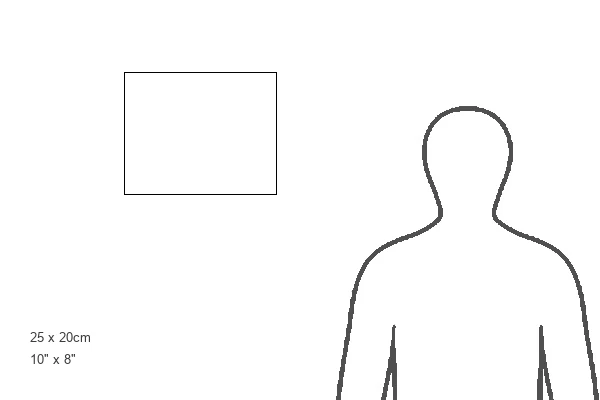
Estimated Product Size is 25.4cm x 20.3cm (10" x 8")
These are individually made so all sizes are approximate
Artwork printed orientated as per the preview above, with landscape (horizontal) or portrait (vertical) orientation to match the source image.
EDITORS COMMENTS
This print showcases the intricate world of nerve and glial cells, captured through a fluorescence light micrograph. The neural stem cells in this image have undergone differentiation, transforming into neurons (nerve cells) depicted in blue, as well as glial cells (support cells) shown in red. The dendrites extending from the neurons are responsible for transmitting electrical signals throughout the nervous system. To enhance visibility and highlight specific proteins, fluorescent markers were employed during sample preparation. In this particular image, beta III-tubulin is stained blue to emphasize its presence within neuronal cytoskeletons. Meanwhile, GFAP (glial fibrillary acidic protein), which forms the structural framework of glial cells' cytoskeletons, appears vibrant red. It's important to note that this sample originates from rat tissue and offers valuable insights into cellular biology and neurology research. By studying these fundamental building blocks of our nervous system – astrocytes, neurones, differentiated stem cells – scientists gain a deeper understanding of how our brains function. The photographer behind this remarkable image is Daniel Schroen from Cell Applications Inc/Science Photo Library. This visually striking photograph not only captures the beauty found within biological structures but also serves as a testament to human curiosity and scientific exploration in unraveling the mysteries of life itself.
MADE IN THE USA
Safe Shipping with 30 Day Money Back Guarantee
FREE PERSONALISATION*
We are proud to offer a range of customisation features including Personalised Captions, Color Filters and Picture Zoom Tools
SECURE PAYMENTS
We happily accept a wide range of payment options so you can pay for the things you need in the way that is most convenient for you
* Options may vary by product and licensing agreement. Zoomed Pictures can be adjusted in the Basket.