
Nervous System Collection
The nervous system, a complex network of pathways and cells, is the powerhouse behind our every move


All Professionally Made to Order for Quick Shipping
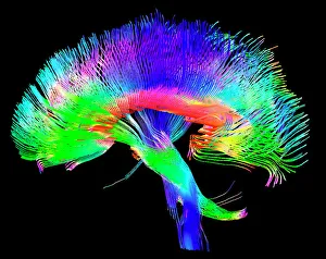
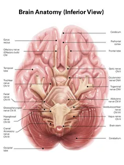
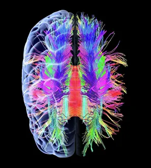
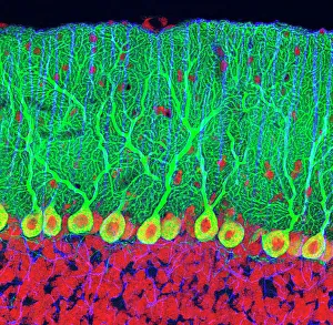
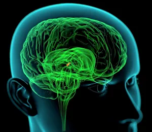
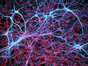
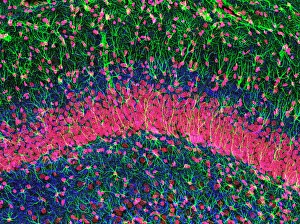
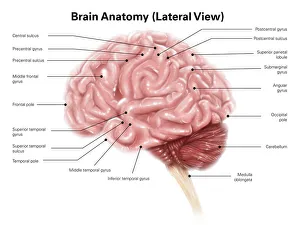
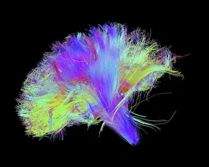
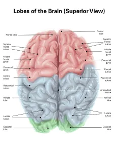
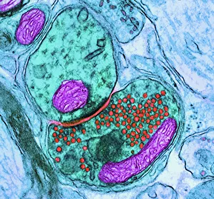
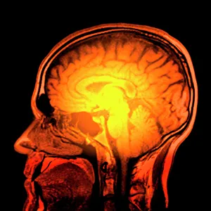
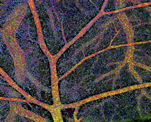
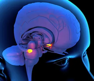
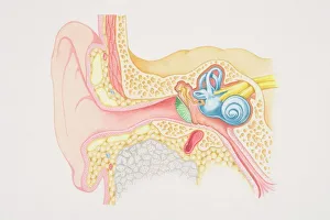
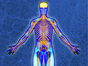
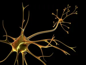
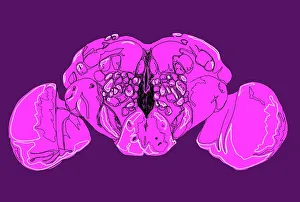
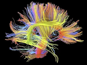
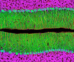
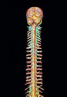
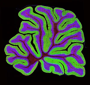
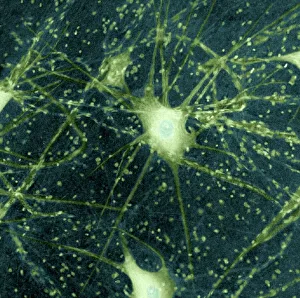
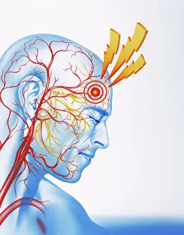
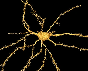
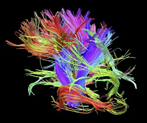
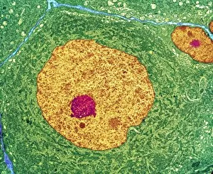
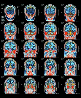
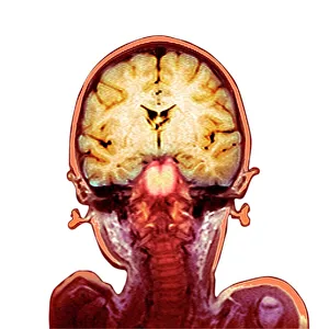
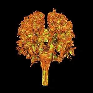
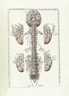
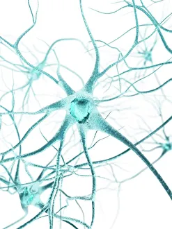
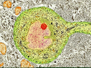
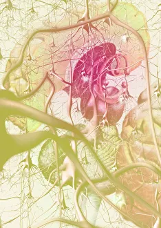
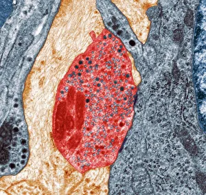
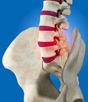
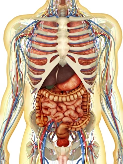
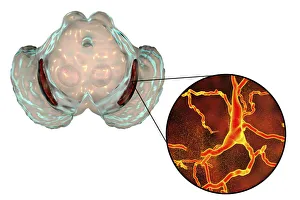
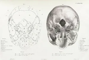
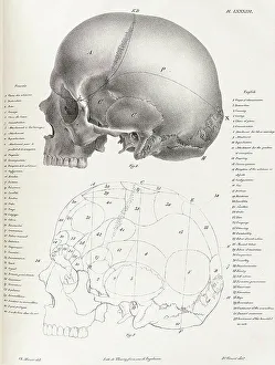
The nervous system, a complex network of pathways and cells, is the powerhouse behind our every move. From the anatomy of the human brain, as seen from an inferior view, to the intricate white matter fibers that connect different regions of this remarkable organ, it's a masterpiece of nature's design. In artwork C015 from 1930, we catch a glimpse of the medulla oblongata nestled within the brain. This vital structure controls essential functions like breathing and heart rate. And speaking of structures, nerve and glial cells are captured in stunning detail under a light microscope - their intricate connections forming the foundation for communication within our nervous system. At synapse nerve junctions depicted through TEM imaging, we witness tiny gaps where signals jump between neurons - an awe-inspiring display of coordination. A superior view showcases colored lobes with labels on a human brain; each lobe responsible for specific functions such as memory or motor control. Moving laterally along this incredible organ's surface reveals its complexity even further. The hippocampus stands out among other brain tissues due to its crucial role in memory formation and spatial navigation. Meanwhile, cross-section diagrams unveil how our ears play an integral part in transmitting sound waves into electrical signals that can be interpreted by our brains. Purkinje nerve cells residing in the cerebellum steal attention with their unique shape and function - fine-tuning movements and maintaining balance effortlessly. Finally, modern technology allows us to explore brain anatomy through MRI scans; unveiling hidden mysteries layer by layer. The nervous system is truly captivating: orchestrating thoughts, sensations, emotions - everything that makes us who we are. Its intricacies continue to astound scientists as they strive to unravel its secrets while appreciating its beauty at every turn.