
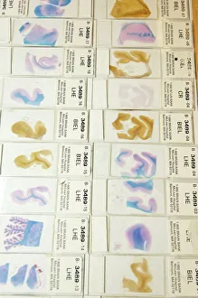
Brain Collection
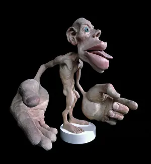
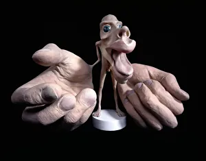
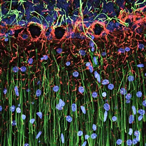
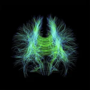
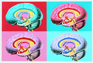
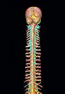
"The intricate map of the brain: Exploring its motor and sensory pathways" Delving into the depths of our mind, we uncover the wonders of the brain's complex anatomy


All Professionally Made to Order for Quick Shipping
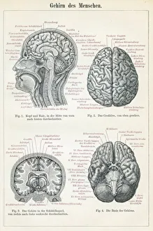
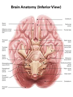
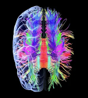
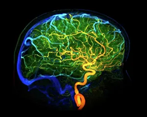
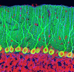
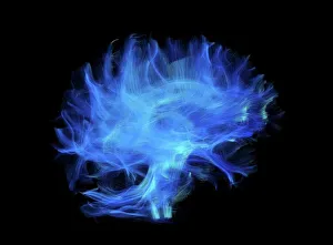
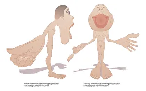
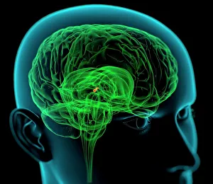
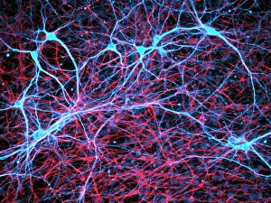
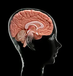
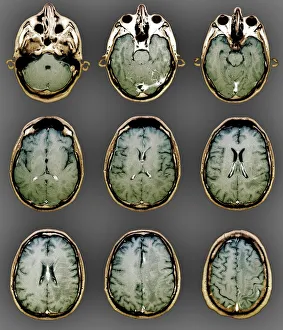
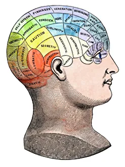
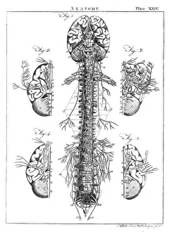
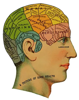
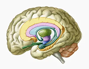
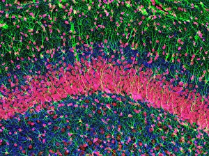
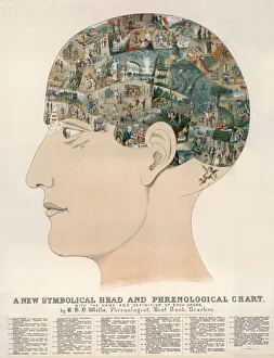
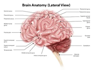
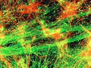
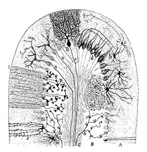
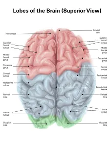
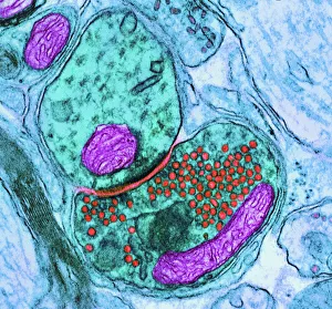
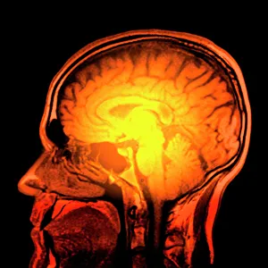
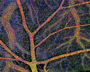
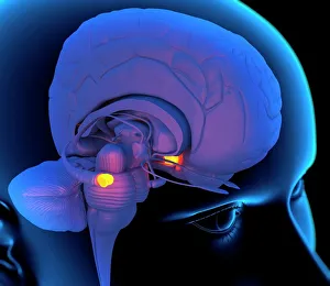
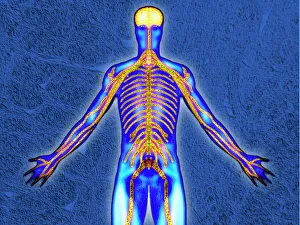
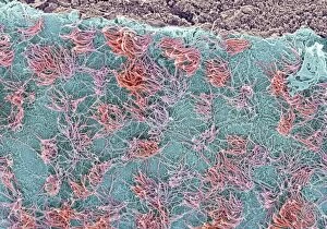
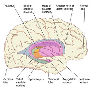
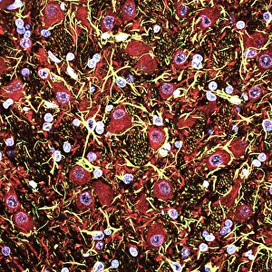
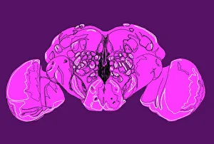
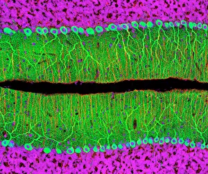
"The intricate map of the brain: Exploring its motor and sensory pathways" Delving into the depths of our mind, we uncover the wonders of the brain's complex anatomy. The Motor homunculus model reveals a distorted representation of our body, with certain regions magnified to depict their significance in movement control. Similarly, the Sensory homunculus showcases how different areas are dedicated to processing various sensations, emphasizing their importance in perceiving touch, pain, and temperature. Transporting us back in time, an engraving from 1895 unveils a vintage depiction anatomy. This historical artwork reminds us of the continuous quest for understanding this enigmatic organ that has captivated scientists for centuries. Zooming closer under a microscope lens, we witness the delicate beauty within cerebellum tissue through a light micrograph. Its intricate structure plays a crucial role in coordinating movements and maintaining balance. The Brain fibres captured by DTI MRI scan C017 / 7099 provide insight into its connectivity network. These fibers act as highways transmitting information between different regions - like bridges connecting distant lands. An inferior view offers another perspective on human brain anatomy. It unravels hidden complexities beneath our surface thoughts – showcasing structures responsible for memory formation, language processing, and emotional regulation. Artistic imagination breathes life into white matter fibres and brains through artwork C015 / 1930. This captivating portrayal merges science with creativity to portray both scientific accuracy and aesthetic appeal simultaneously. Venturing deeper within this mysterious realm is an exploration blood vessels using a stunning 3D angiogram C007 /1981. These intricate networks supply oxygen-rich blood to nourish every nook and cranny of this vital organ. Returning once again to explore motor and sensory functions intricately mapped out by homunculi models – these visual representations highlight how specific regions control precise movements or perceive distinct sensations throughout our body A masterpiece artwork captures Medulla oblongata in the brain, showcasing its vital role in controlling essential functions