
Tubulin Collection
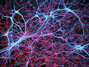
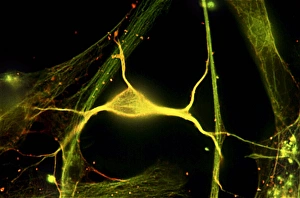
Tubulin: The Backbone of Cellular Infrastructure Nerve and glial cells, as seen under a light micrograph, are intricately connected by tubulin


All Professionally Made to Order for Quick Shipping
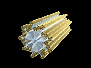
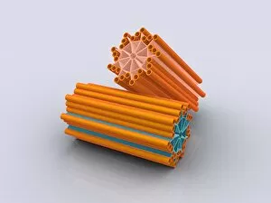
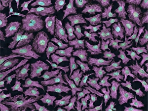
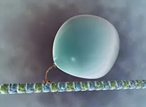
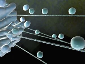
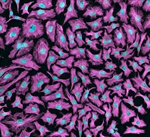
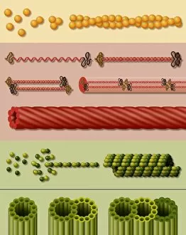
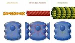
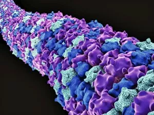
Tubulin: The Backbone of Cellular Infrastructure Nerve and glial cells, as seen under a light micrograph, are intricately connected by tubulin, a vital protein that forms microtubules within the cell. These microscopic structures act as highways for intracellular transport and provide structural support. In an artistic representation, the interplay between tubulin and microtubules is beautifully depicted. This artwork showcases how this protein orchestrates the intricate dance of cellular activities, ensuring proper functioning and organization. A conceptual image of centriole highlights its crucial role in cell division. Tubulin plays a pivotal part in organizing these spindle-shaped structures during mitosis to ensure accurate chromosome segregation. Multiphoton fluorescence images of HeLa cells reveal the dynamic nature of tubulin-mediated processes within living cells. These images capture the vibrant glow emitted by fluorescently labeled tubulin as it forms complex networks throughout the cytoplasm. Illustrations depicting microtubule formation showcase how tubulin molecules assemble into long hollow tubes with remarkable precision. This process is essential for maintaining cell shape, facilitating intracellular transport, and enabling various cellular functions. Artwork illustrating intracellular transport demonstrates how tubulin acts as a conveyor belt inside cells. It transports vital cargo such as organelles and vesicles to their designated locations with utmost efficiency and accuracy. HeLa cells captured under a light microscope exhibit the intricate web-like network formed by tubulin filaments. This visual representation emphasizes its critical role in maintaining cellular architecture while providing stability to these rapidly dividing cancerous cells.