Fraud Blocker Skip to main content

Neuron Collection

"Unveiling the Intricacies of Neurons: Exploring the Wonders Within Our Brain" Delving into the depths of our brain

Background imageNeuron Collection: Histological Diagram of a Mammalian Retina

Histological Diagram of a Mammalian Retina
RAMON Y CAJAL, Santiago (1852-1934). Spanish doctor and histologist, Nobel Prize in 1906. Histological diagram of a mammalian retina. Original drawing by Ramon y Cajal

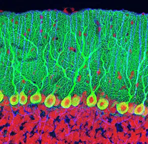
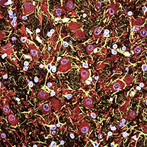
Background imageNeuron Collection: Cerebellum tissue, light micrograph

Cerebellum tissue, light micrograph
Cerebellum tissue. Confocal light micrograph of a section through the cerebellum of the brain. Purkinje cells, a type of neuron (nerve cell), are red

Background imageNeuron Collection: Purkinje nerve cells in the cerebellum

Purkinje nerve cells in the cerebellum
Purkinje cells in the cerebellum. Fluorescent light micrograph of Purkinje cells (green) in the cerebellum of the brain. Purkinje nerve cells have a flask-like body from which numerous highly

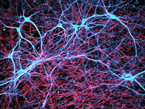
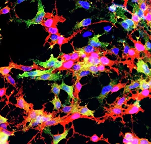
Background imageNeuron Collection: Nerve and glial cells, light micrograph

Nerve and glial cells, light micrograph
Nerve and glial cells, fluorescence light micrograph. These are neural stem cells that have differentiated into neurons (nerve cells, blue) and glial cells (support cells, red)

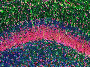
Background imageNeuron Collection: Hippocampus brain tissue

Hippocampus brain tissue
Hippocampus tissue. Light micrograph of a sagittal (side view) section through the hippocampus of the brain showing the nerve cells within it

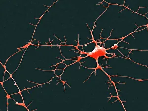
Background imageNeuron Collection: Nerve cell, SEM

Nerve cell, SEM
Nerve cell. Coloured scanning electron micrograph (SEM) of a nerve cell (neuron). Neurons are responsible for passing information around the central nervous system (CNS)

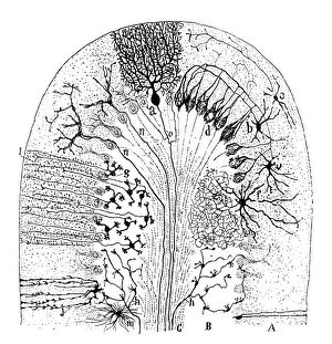
Background imageNeuron Collection: Cell types in the mammalian cerebellum: drawing, 1894, by the Spanish histologist Santiago Ramon y

Cell types in the mammalian cerebellum: drawing, 1894, by the Spanish histologist Santiago Ramon y Cajal (1852-1934)
NERVE CELLS, 1894. Cell types in the mammalian cerebellum: drawing, 1894, by the Spanish histologist Santiago Ramon y Cajal (1852-1934)

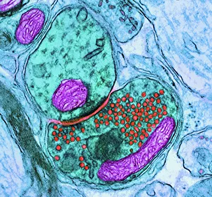
Background imageNeuron Collection: Synapse nerve junction, TEM

Synapse nerve junction, TEM
Synapse. Coloured transmission electron micrograph (TEM) of a synapse, a junction between two nerve cells, in the brain. At a synapse an electrical signal is transmitted from one cell to the next in

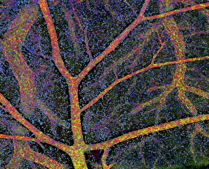
Background imageNeuron Collection: Brain tissue blood supply

Brain tissue blood supply. Light micrograph of a section through cortex tissue from a brain, showing the blood vessels (branching) that supply it

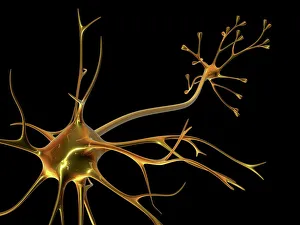
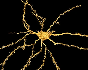
Background imageNeuron Collection: Nerve cell

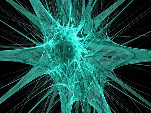
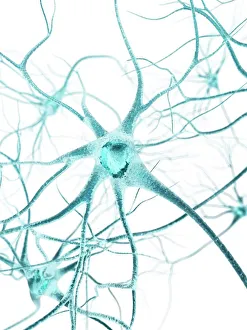
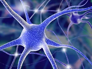
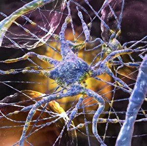
Nerve cell. Computer artwork of a nerve cell, also called a neuron. Neurons are responsible for passing information around the central nervous system (CNS) and from the CNS to the rest of the body

Background imageNeuron Collection: Glial stem cell culture, light micrograph

Glial stem cell culture, light micrograph
Glial stem cell culture. Fluorescent light micrograph of glial stem cells producing the protein NG2 (red) as they mature. These stem cells can differentiate into several types of glial cells

Background imageNeuron Collection: Cerebral cortex nerve cells

Cerebral cortex nerve cells. Confocal light micrograph of neurons (nerve cells, red) and glial cells (support cells, gold) from the cerebral cortex

Background imageNeuron Collection: Purkinje nerve cells in the cerebellum

Purkinje nerve cells in the cerebellum
Purkinje cells in the cerebellum. Fluorescent light micrograph of Purkinje cells (green) in the cerebellum of the brain. Purkinje nerve cells have a flask-like body from which numerous highly
Background imageNeuron Collection: Myelination of nerve fibres, TEM

Myelination of nerve fibres, TEM
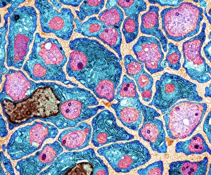
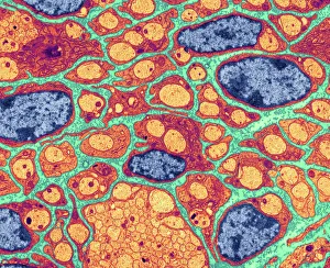
Myelination of nerve fibres. Coloured transmission electron micrograph (TEM) of Schwann cells (blue, with brown nuclei) insulating nerve fibres (axons, pink) with a myelin sheath

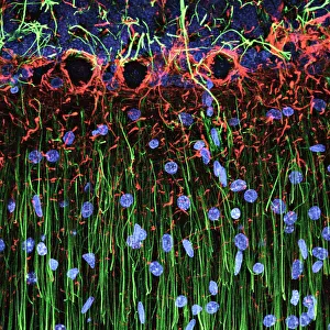
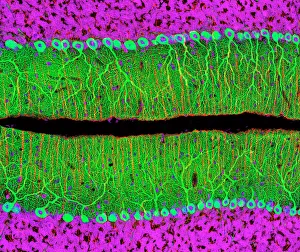
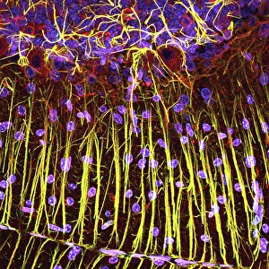
Background imageNeuron Collection: Cerebellum structure, light micrograph

Cerebellum structure, light micrograph
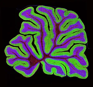
Cerebellum structure. Fluorescent light micrograph of a section through the cerebellum of the brain. The cerebellum comprises three main layers

Background imageNeuron Collection: Neural stem cell culture

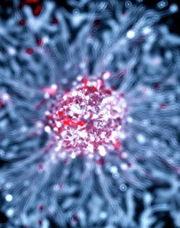
Neural stem cell culture. Fluorescent light micrograph of a group of neural stem cells (neurosphere) in culture. Neural stem cells are able to differentiate into neurons (nerve cells)

Background imageNeuron Collection: Motor neurons, light micrograph

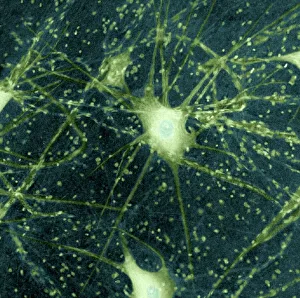
Motor neurons, light micrograph. Motor neurons are responsible for passing information around the central nervous system (CNS) and from the CNS to the rest of the body

Background imageNeuron Collection: Brain neuron

Brain neuron. Computer reconstruction of a medium spiny neuron from the basal ganglia of the brain. Neurons (nerve cells) are responsible for passing information around the central nervous system

Background imageNeuron Collection: Cerebellum tissue, light micrograph

Cerebellum tissue, light micrograph
Cerebellum tissue. Confocal light micrograph of a section through the cerebellum of the brain. Purkinje cells, a type of neuron (nerve cell), are red

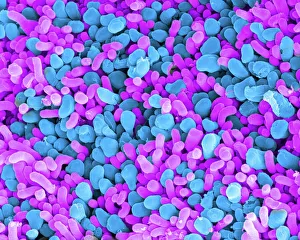
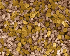
Background imageNeuron Collection: Rod and cone cells of the eye, SEM C014 / 4866

Rod and cone cells of the eye, SEM C014 / 4866
Rod and cone cells of the eye. Coloured scanning electron micrograph (SEM) of rod and cone cells in the retina of a mammalian eye

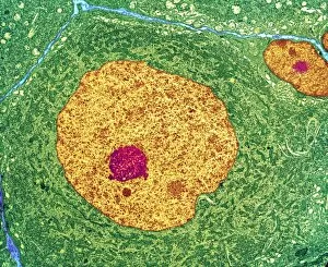
Background imageNeuron Collection: Nerve cell, TEM

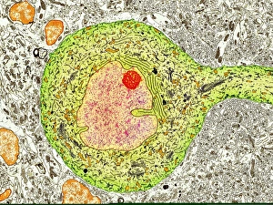
Nerve cell, TEM
Nerve cell. Coloured transmission electron micrograph (TEM) of a nerve cell body in cross- section. The cell has a large nucleus (yellow) and inner nucleolus (red)

Background imageNeuron Collection: Nerve cells, abstract artwork

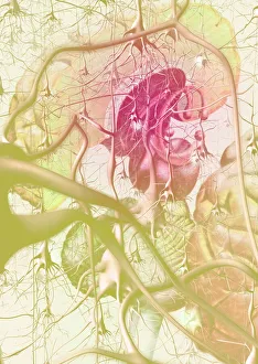
Nerve cells, abstract artwork
Nerve cells. Abstract computer artwork of nerve cells, or neurons. Neurons are responsible for passing information around the central nervous system (CNS) and from the CNS to the rest of the body

Background imageNeuron Collection: Myelination of nerve fibres, TEM

Myelination of nerve fibres, TEM
Myelination of nerve fibres. Coloured transmission electron micrograph (TEM) of Schwann cells (red, with blue nuclei) insulating nerve fibres (axons, orange) with a myelin sheath

Background imageNeuron Collection: Myelinated nerve, TEM

Myelinated nerve, TEM
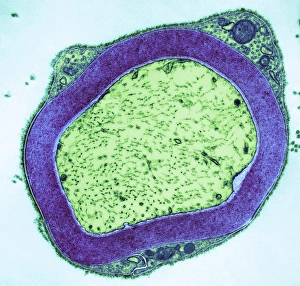
Myelinated nerve. Coloured transmission electron micrograph (TEM) of myelinated nerve fibres and Schwann cells. Myelin (purple) is an insulating fatty layer that surrounds nerve fibres (axons)

Background imageNeuron Collection: Synapse nerve junctions, SEM

Synapse nerve junctions, SEM
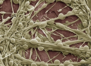
Synapse nerve junctions. Coloured scanning electron micrograph (SEM) of nerve cells showing the synapses (junctions, bulges) between them

Background imageNeuron Collection: Nerve cell, artwork F007 / 7448

Nerve cell, artwork F007 / 7448
Nerve cell, computer artwork

Background imageNeuron Collection: Purkinje nerve cell, TEM C014 / 0583

Purkinje nerve cell, TEM C014 / 0583
Purkinje nerve cell. Transmission electron micrograph (TEM) of a purkinje nerve cell (green) from the cerebellum of the brain, showing the cell body (centre) and its primary dendrite (cell process)

Background imageNeuron Collection: Brain anatomy

Brain anatomy. Computer artwork of strands of nerve cells (neurons) in front of an exploded view of the brain seen from behind

Background imageNeuron Collection: Nerve synapse, TEM

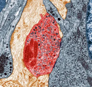
Nerve synapse, TEM
Nerve synapse. Coloured transmission electron micrograph (TEM) of the neuron (nerve) terminal at a synapse in the diaphragm

Background imageNeuron Collection: Nerve cell, SEM

Nerve cell, SEM
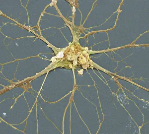
Nerve cell. Coloured scanning electron micrograph (SEM) of a neuron (nerve cell). The cell body is the central structure with neurites (long and thin structures) radiating outwards from it

Background imageNeuron Collection: Nerve cells, computer artwork

Nerve cells, computer artwork
Nerve cells. Computer artwork of nerve cells, or neurons

Background imageNeuron Collection: Map of nerve bundles in the human brain by Paul Broca

Map of nerve bundles in the human brain by Paul Broca
5309460 Map of nerve bundles in the human brain by Paul Broca; (add.info.: Map of nerve bundles in the human brain by Paul Broca)

Background imageNeuron Collection: An engraving depicting a diagram showing the supposed functions of various areas of the brain

An engraving depicting a diagram showing the supposed functions of various areas of the brain, 19th century
5311364 An engraving depicting a diagram showing the supposed functions of various areas of the brain, 19th century; (add.info.: Date: 1886); Universal History Archive/UIG.

Background imageNeuron Collection: An engraving depicting Descartes' drawing of the human brain, 17th century

An engraving depicting Descartes' drawing of the human brain, 17th century
5311365 An engraving depicting Descartes' drawing of the human brain, 17th century; Universal History Archive/UIG.

Background imageNeuron Collection: Wilhelm Waldeyer, 1880

Wilhelm Waldeyer, 1880
1576978 Wilhelm Waldeyer, 1880; (add.info.: Wilhelm Waldeyer (6th October 1836 - 23 January 1921) was a German anatomist, famous for development of the neuron theory.); Universal History Archive/UIG.

Background imageNeuron Collection: A cartoon commenting on the fashionable revival of phrenology, 19th century

A cartoon commenting on the fashionable revival of phrenology, 19th century
5311366 A cartoon commenting on the fashionable revival of phrenology, 19th century; (add.info.: Date: 1878); Universal History Archive/UIG.

Background imageNeuron Collection: Neuron

Neuron
Mei Xu

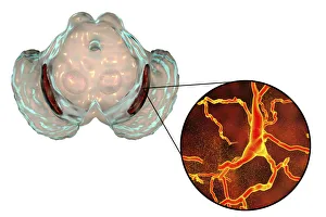
Background imageNeuron Collection: Substantia nigra in Parkinsons disease, illustration

Substantia nigra in Parkinsons disease, illustration
Substantia nigra. Computer illustration showing a degenerated substantia nigra and dopaminergic neurons in Parkinsons disease

Background imageNeuron Collection: Ramon y Cajal, Santiago (Petilla of Aragon, Navarra, 1852-Madrid, 1934)

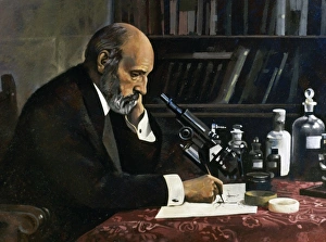
Ramon y Cajal, Santiago (Petilla of Aragon, Navarra, 1852-Madrid, 1934). Professor of Medicine and Surgery. Spanish histologist and pathologist

Background imageNeuron Collection: Santiago Ramon y Cajal (Petilla of Aragon, Navarra, 1852-Madrid, 1934), Professor

Santiago Ramon y Cajal (Petilla of Aragon, Navarra, 1852-Madrid, 1934), Professor of Medicine and Surgery, histologist and pathologist

Background imageNeuron Collection: Santiago Ramon and Cajal (1852-1934) Spanish histologist, physician and pathologist

Santiago Ramon and Cajal (1852-1934) Spanish histologist, physician and pathologist
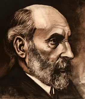
Santiago Ramon and Cajal (1852-1934).. Spanish histologist, physician and pathologist. He made important discoveries such as laws governing the morphology and connections of nerve cells in the brain

Background imageNeuron Collection: brain cells

brain cells
microscopic image

Background imageNeuron Collection: ai, ar, artificial, artificial intelligence, atom, augmented reality, beauty, brain

ai, ar, artificial, artificial intelligence, atom, augmented reality, beauty, brain
Woman with artificial intelligence brain

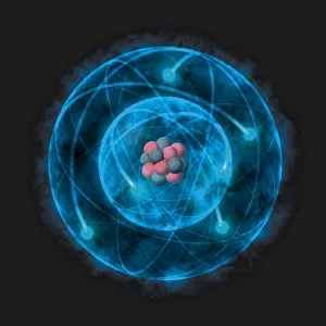
Background imageNeuron Collection: Illustration of atom with nucleus of protons and neurons, based on the Bohr model

Illustration of atom with nucleus of protons and neurons, based on the Bohr model

Background imageNeuron Collection: Transverse section of the midbrain

Transverse section of the midbrain

Background imageNeuron Collection: Nerve synapse and serotonin molecule

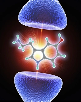
Nerve synapse and serotonin molecule
Nerve synapse and serotonin neurotransmitter molecule. Computer artwork of a junction, or synapse, between two nerve cells (neurons, blue)

Background imageNeuron Collection: Neurology slide show C016 / 9008

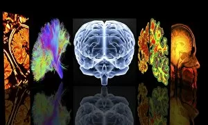
Neurology slide show C016 / 9008
Conceptual computer artwork depicting neurology. From left to right: MRI brain scans, 3D dsi white matter brain scan, brain, Alzheimers brain versus normal brain, MRI brain scan

Background imageNeuron Collection: Rod and cone cells of the eye, SEM C014 / 4864

Rod and cone cells of the eye, SEM C014 / 4864
Rod and cone cells of the eye. Coloured scanning electron micrograph (SEM) of rod and cone cells in the retina of a mammalian eye


All Professionally Made to Order for Quick Shipping

"Unveiling the Intricacies of Neurons: Exploring the Wonders Within Our Brain" Delving into the depths of our brain, we encounter a histological diagram of a mammalian retina. This intricate network showcases the complexity and beauty of neurons that enable us to perceive light and color. Moving further, we explore cerebellum tissue through a light micrograph. The mesmerizing patterns reveal nerve and glial cells working in harmony, orchestrating our body's movements with precision. Zooming in closer, we witness a synapse nerve junction captured by TEM. This microscopic marvel highlights how information is transmitted between neurons, forming connections crucial for our thoughts and actions. Shifting gears to SEM imagery, we are introduced to an awe-inspiring nerve cell. Its intricate structure resembles an elaborate work of art—a testament to nature's ingenuity in crafting these building blocks of intelligence. Tracing back history, we stumble upon Santiago Ramon y Cajal's 1894 drawing depicting cell types within the mammalian cerebellum. His meticulous observations laid foundations for understanding neural networks that govern our motor skills. Venturing deeper into brain tissue, we discover hippocampus tissue—an essential region responsible for memory formation and spatial navigation. Here lies another realm where neurons weave together memories that shape who we are. Intriguingly unique are Purkinje nerve cells found within the cerebellum—majestic giants among their peers. Their distinctive appearance signifies their vital role in coordinating movement and maintaining balance. As if peering through a microscope lens once again, another nerve cell captures our attention—the epitome of elegance amidst complexity; it reminds us how intricately woven life truly is at its core. Diving into glial stem cell culture under bright illumination reveals their remarkable regenerative potential—a beacon of hope for treating neurological disorders as they hold promises yet untapped. Examining brain tissue blood supply uncovers an indispensable lifeline, nourishing neurons with oxygen and nutrients.