Photographic Print : Nerve ganglion, light micrograph
![]()

Photo Prints from Science Photo Library
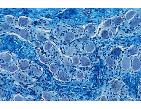
Nerve ganglion, light micrograph
Nerve ganglion. Light micrograph of a section through a dorsal (sensory) spinal root ganglion associated with a sensory nerve root of the spinal cord. Sensory information from peripheral sites e.g. skin, muscle, organs enters the spinal cord via the axons of dorsal roots. The nerve cell bodies of dorsal root axons are clustered in a ganglion which is noted as a discrete swelling of the nerve root. The large spherical cell bodies have a single nucleus and each body is surrounded by glial/satellite cells. Axons between the cell bodies are myelinated with Schwann cells. Dorsal root ganglia are classed as pseudounipolar nerve cells because they have no dendrites; the afferent and efferent nerve fibres linked to each cell body are axons. Magnification: x65
Science Photo Library features Science and Medical images including photos and illustrations
Media ID 9247137
© MICROSCAPE/SCIENCE PHOTO LIBRARY
Axon Axons Bodies Cell Biology Cell Body Central Nervous System Cytological Cytology Dorsal Fibre Fibres Ganglion Glial Cell Histological Histology Myelin Myelinated Nerve Nerves Neuron Neurone Neurones Neurons Neuroscience Nuclei Nucleus Schwann Cell Sensory Sheath Sheaths Spinal Cord Stain Stained Swelling Tissue Cells Light Micrograph Light Microscope Section Sectioned
11"x8.5" Photo Print
Discover the beauty and complexity of the natural world with Media Storehouse's range of Photographic Prints. This captivating image, taken from the Science Photo Library, showcases a light micrograph of a nerve ganglion. Witness the intricate details of a dorsal (sensory) spinal root ganglion, associated with a sensory nerve root of the spinal cord. Each print is produced with the highest quality standards, ensuring vibrant colors and sharp details that bring the wonders of science right into your home or office. Order now and let this stunning image inspire your curiosity and deepen your understanding of the world around us.
Photo prints are produced on Kodak professional photo paper resulting in timeless and breath-taking prints which are also ideal for framing. The colors produced are rich and vivid, with accurate blacks and pristine whites, resulting in prints that are truly timeless and magnificent. Whether you're looking to display your prints in your home, office, or gallery, our range of photographic prints are sure to impress. Dimensions refers to the size of the paper in inches.
Our Photo Prints are in a large range of sizes and are printed on Archival Quality Paper for excellent colour reproduction and longevity. They are ideal for framing (our Framed Prints use these) at a reasonable cost. Alternatives include cheaper Poster Prints and higher quality Fine Art Paper, the choice of which is largely dependant on your budget.
Estimated Image Size (if not cropped) is 27.9cm x 18.4cm (11" x 7.2")
Estimated Product Size is 27.9cm x 21.6cm (11" x 8.5")
These are individually made so all sizes are approximate
Artwork printed orientated as per the preview above, with landscape (horizontal) orientation to match the source image.
EDITORS COMMENTS
This print showcases a nerve ganglion in all its intricate glory. Taken under a light microscope, this image reveals a section through a dorsal spinal root ganglion associated with a sensory nerve root of the spinal cord. The ganglion, characterized by its discrete swelling, houses the nerve cell bodies of dorsal root axons. The large spherical cell bodies take center stage in this composition, each containing a single nucleus and surrounded by glial/satellite cells. Connecting these cell bodies are myelinated axons, sheathed with Schwann cells. It is fascinating to observe how sensory information from peripheral sites such as skin, muscle, and organs enters the spinal cord via these axons. Dorsal root ganglia are classified as pseudounipolar nerve cells due to their lack of dendrites; instead, both the afferent and efferent nerve fibers linked to each cell body function as axons. This microscopic view offers us an insight into the complex network that enables our sense of touch and perception. With magnification at x65, this image not only highlights the beauty within our biological makeup but also serves as an invaluable resource for researchers delving into fields like neuroscience and histology. By capturing this moment in time using advanced staining techniques on healthy tissue samples, Science Photo Library provides us with yet another remarkable glimpse into the wonders of human anatomy at its cellular level.
MADE IN THE USA
Safe Shipping with 30 Day Money Back Guarantee
FREE PERSONALISATION*
We are proud to offer a range of customisation features including Personalised Captions, Color Filters and Picture Zoom Tools
SECURE PAYMENTS
We happily accept a wide range of payment options so you can pay for the things you need in the way that is most convenient for you
* Options may vary by product and licensing agreement. Zoomed Pictures can be adjusted in the Cart.


