Home > Popular Themes > Human Body
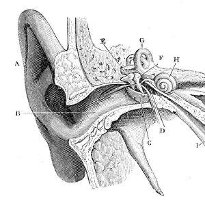
The human ear, wood engraving, published in 1880

![]()
Wall Art and Photo Gifts from Fine Art Storehouse
The human ear, wood engraving, published in 1880
Anatomy of the human ear: A) auricle, B) External Auditory Canal, C) Tympanic Membrane, D) Tympanic Cavity, E) Malleus, M) Incus, H) Cochlea, G) Semicircular Canals, I) Eustachian Tube. Wood engraving, published in 1880
Unleash your creativity and transform your space into a visual masterpiece!
ZU_09
Media ID 11772532
© ZU_09
78279 Anatomic Anatomy Biomedical Illustration Body Part Cochlea Ear Drum Eustachian Tube Human Ear Human Internal Organ Image Date Incus Listening Malleus Middle Ear Physiology Semicircular Canal Tympanic Cavity Wood Engraving Earlobe Human Body Part Inner Ear Internal System
FEATURES IN THESE COLLECTIONS
> Fine Art Storehouse
> The Magical World of Illustration
> Art Illustrations
EDITORS COMMENTS
This wood engraving, published in 1880, offers a detailed glimpse into the intricate anatomy of the human ear. The print showcases various components of this remarkable sensory organ, providing an invaluable resource for understanding its structure and function. Starting from the outermost part, we see the auricle (A), which acts as a funnel to collect sound waves. Moving inward, we encounter the external auditory canal (B) leading to the tympanic membrane (C), commonly known as the eardrum. This delicate membrane separates the outer ear from the middle ear. Within the middle ear lies an array of vital structures depicted in this engraving: The tympanic cavity (D), malleus (E), incus (M), and cochlea (H). These elements work harmoniously to transmit sound vibrations from air to fluid-filled chambers within our inner ears. The semicircular canals (G) are also visible in this illustration, responsible for maintaining balance and spatial orientation. Lastly, we observe the Eustachian tube (I), connecting our ears with our throat and playing a crucial role in equalizing pressure between our middle ears and atmospheric conditions. This historic artwork not only serves as a testament to scientific advancements during that era but also highlights humanity's enduring fascination with unraveling nature's mysteries. It invites us to appreciate both artistry and knowledge by capturing a moment frozen in time – forever preserving our understanding of one of life's most extraordinary senses.
MADE IN THE USA
Safe Shipping with 30 Day Money Back Guarantee
FREE PERSONALISATION*
We are proud to offer a range of customisation features including Personalised Captions, Color Filters and Picture Zoom Tools
SECURE PAYMENTS
We happily accept a wide range of payment options so you can pay for the things you need in the way that is most convenient for you
* Options may vary by product and licensing agreement. Zoomed Pictures can be adjusted in the Cart.