
Ear Drum Collection
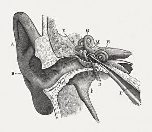
"The Intricate Symphony: Exploring the Delicate Mechanism of the Ear" Step back in time to 1876 with a captivating lithograph showcasing the anatomy of the human ear


All Professionally Made to Order for Quick Shipping
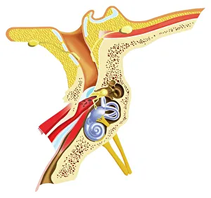
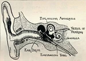
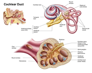
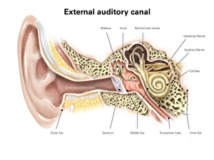
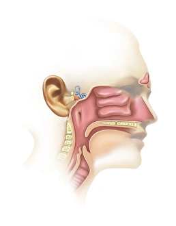
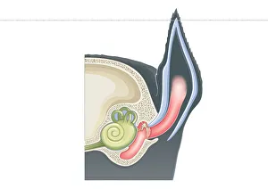
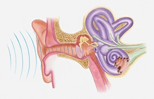
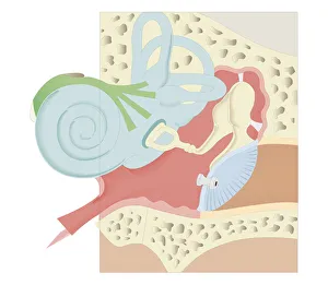
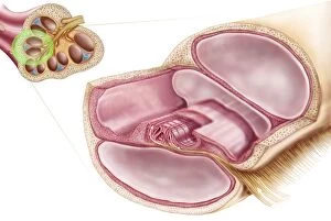
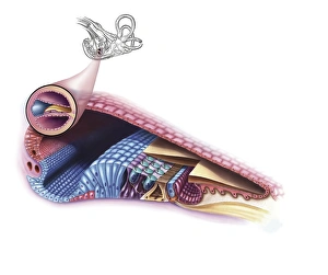
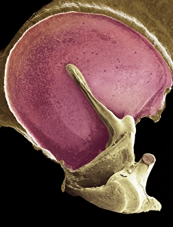
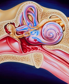
"The Intricate Symphony: Exploring the Delicate Mechanism of the Ear" Step back in time to 1876 with a captivating lithograph showcasing the anatomy of the human ear. This vintage masterpiece takes us on a journey through the intricate inner workings of this remarkable sensory organ. A detailed diagram reveals the wonders hidden within our auditory canal, including the eardrum, semicircular canals, cochlea, cochlear nerve, and even the eustachian tube. Each element plays a vital role in capturing sound waves and transforming them into meaningful sensations. The Wagner/Gill/L eclipse captures our attention as we marvel at how such a delicate structure can orchestrate our ability to hear. It reminds us that nature's design is truly awe-inspiring. Fast forward to 1934 when "Delicate Mechanism of the Ear" was brought to life through another stunning illustration. The complexity of this system becomes more apparent as we explore every nook and cranny of its inner workings. The cochlear duct takes center stage in one image, emphasizing its significance in translating vibrations into electrical signals for our brain to interpret. Meanwhile, an external auditory canal diagram provides labels that guide us through each component's purpose. Venturing further into this captivating realm brings us face-to-face with both sinuses and inner ear anatomy. These illustrations serve as reminders that hearing is not just limited to sound but also encompasses balance and spatial awareness – all made possible by this extraordinary creation. A cutaway diagram offers an intriguing glimpse into what lies beneath our skin – revealing layers upon layers intricately connected like Uppsala Cathedral soaring high above Scandinavia's skyline. Just like this architectural marvel stands tall amidst its surroundings, so does our ear amidst other organs within our body. Finally, modern digital illustrations bring clarity to mammalian ears' cross-sections while highlighting their pinnae (external ears), eardrums, and middle ear structures.