
Malleus Collection
"Malleus: Unveiling the Intricacies of the Human Ear through Scientific Illustrations" Step back in time to 1876 with this captivating lithograph


All Professionally Made to Order for Quick Shipping
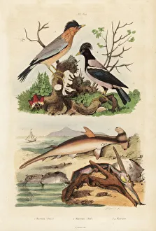
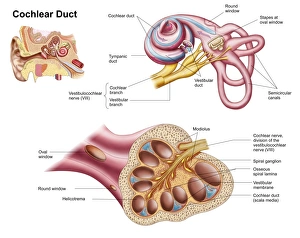
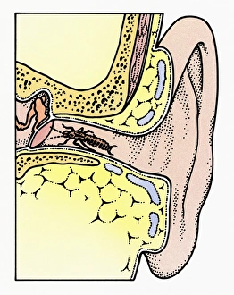
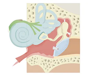
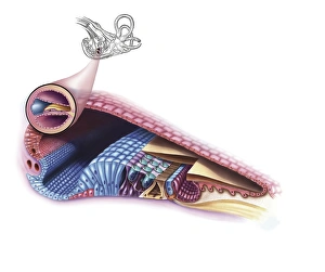
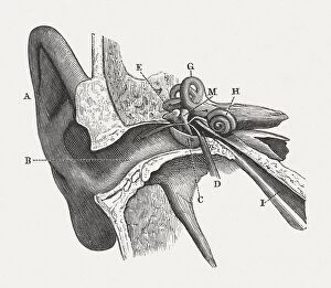
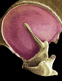
"Malleus: Unveiling the Intricacies of the Human Ear through Scientific Illustrations" Step back in time to 1876 with this captivating lithograph, a masterpiece that delves into the anatomy of the human ear. Published during an era when scientific illustrations were highly valued, this lithograph offers a glimpse into our auditory system like never before. As you explore this intricate artwork, you may be reminded of various references to "malleus" throughout history. From the infamous "Malleus Maleficarum, " a treatise on witchcraft, to its literal translation as "hammer, " it becomes evident why this term is so significant. Marvel at how these detailed illustrations showcase not only the external features but also delve deep within. Discover fascinating connections between different species and their unique adaptations – from the smooth hammerhead shark's distinctive head shape to the hammer oyster's intriguing shell structure. Venturing beyond marine life, observe other molluscs depicted in vibrant colors from around 1860. The cone and wentletrap shells stand out among them, showcasing nature's artistic prowess. But let us not forget our focus on human anatomy. Delve further into these scientific illustrations and witness cross-sections revealing astonishing details about our cochlear duct and external auditory canal. With labels guiding your exploration, unravel each component intricately woven within our ears' complex architecture. Intriguingly enough, even insects find their way into this narrative. Observe a cutaway diagram featuring a common earwig nestled within an auditory canal; its antennae gently touch upon the tympanic membrane—an unexpected yet mesmerizing sight. Finally, immerse yourself in digital artistry depicting mammal middle ears where malleus takes center stage alongside incus and stapes—three tiny bones responsible for transmitting sound waves deep inside our hearing apparatus. This collection of scientific illustrations invites us to appreciate both natural wonders and human ingenuity alike.