
Incus Collection
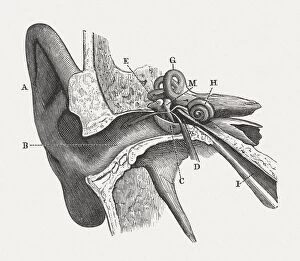
The intricate anatomy of the human ear is beautifully depicted in this lithograph, published in 1876


All Professionally Made to Order for Quick Shipping
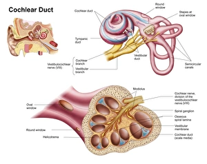
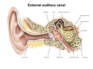
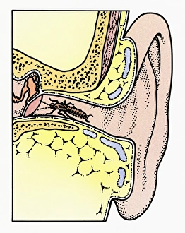
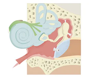
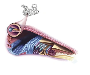
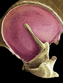
The intricate anatomy of the human ear is beautifully depicted in this lithograph, published in 1876. The scientific illustrations showcase the Ear and Auditory system, providing a detailed look at various components. One such component is the incus, one of the three tiny bones found in the middle ear. In this illustration, we can see a cross-section of the Common Earwig (Forficula auricularia) nestled within the auditory canal of an ear. Its delicate antennae gently touch the tympanic membrane, highlighting how closely connected these structures are. The cutaway diagram offers a comprehensive view of the human ear's internal structure. It reveals not only the cochlear duct but also emphasizes how crucially positioned bones like malleus, incus, and stapes are for our hearing abilities. A digital illustration further highlights these middle ear bones - malleus, incus, and stapes - giving us a close-up view that showcases their unique shapes and functions. These small yet vital elements work together to transmit sound vibrations from our eardrum to our inner ear. Another fascinating biomedical illustration presents a cross-section view of both outer and inner parts of the human ear. This depiction allows us to appreciate how each section contributes to our ability to hear sounds accurately. Lastly, we observe a cross-section biomedical illustration displaying a grommet inserted into an eardrum or tympanic membrane. This medical intervention helps alleviate certain conditions by equalizing pressure within the middle ear space while allowing fluid drainage when necessary. These captivating illustrations remind us just how remarkable our sense of hearing truly is – an intricate collaboration between numerous anatomical structures including those involving incus – enabling us to experience life's symphony with clarity and depth.