
Middle Ear Collection
The middle ear, a fascinating component of the human auditory system, has been depicted in various illustrations throughout history


All Professionally Made to Order for Quick Shipping
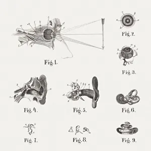
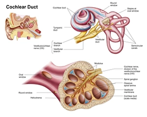
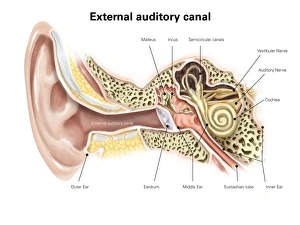
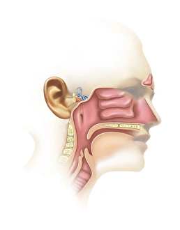
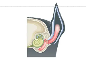
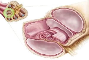
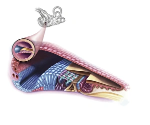
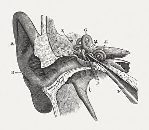
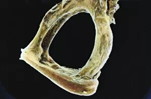
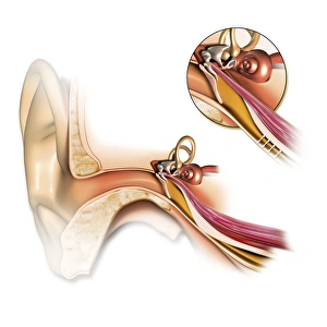
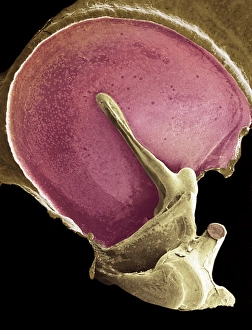
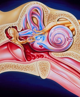
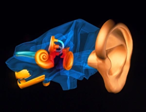
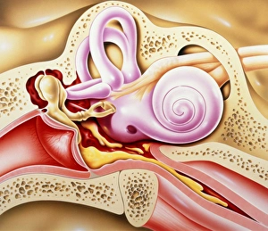
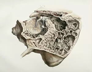
The middle ear, a fascinating component of the human auditory system, has been depicted in various illustrations throughout history. One such lithograph, published in 1876, showcases the intricate anatomy of the human ear. Another notable publication from 1861 provides an insightful depiction of both the eye and ear's anatomy. Among these depictions is an illustration specifically focusing on the cochlear duct within the human ear. This detailed portrayal sheds light on this crucial part responsible for transmitting sound signals to our brain. Another captivating image reveals the external auditory canal of the human ear, complete with labels that help us understand its structure better. Additionally, there are illustrations showcasing both inner ear and sinus anatomy – highlighting their interconnectedness and importance in maintaining balance and equilibrium. A cutaway diagram offers a comprehensive view of how different components work together within our ears. From pinna to eardrum to middle ear structures like ossicles (malleus, incus, stapes), this digital cross-section illustrates mammalian ears' complexity. Furthermore, a digital illustration zooms in on both middle and inner parts of our ears – emphasizing their significance in perceiving sounds accurately. Cross-sectional images further enhance our understanding by depicting all three sections: inner, outer, and middle ears side by side. Lastly but not leastly is a close-up illustration solely dedicated to showcasing middle ear bones - malleus (hammer), incus (anvil), and stapes (stirrup). These tiny yet vital elements play a pivotal role in transmitting sound vibrations from eardrum to cochlea. Even beyond humans' realm lies another intriguing example – an illustrative cross-section displaying domestic cat's (Felis Catus) remarkable feline hearing apparatus. These diverse visual representations offer valuable insights into one aspect we often take for granted - our ability to hear. Exploring these depictions allows us to appreciate just how intricately designed our ears are; enabling us to experience the symphony of sounds that surround us every day.