
Cochlea Collection
The cochlea, an intricate structure nestled within the human ear, has captivated scientists and artists alike throughout history


All Professionally Made to Order for Quick Shipping
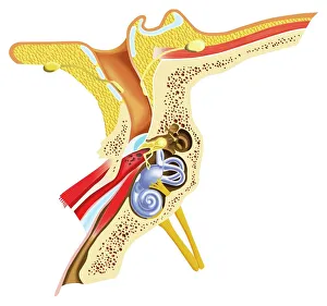
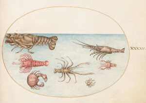
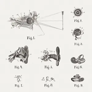
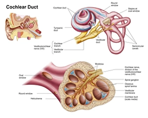
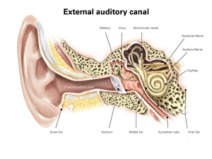
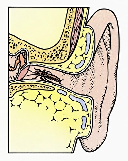
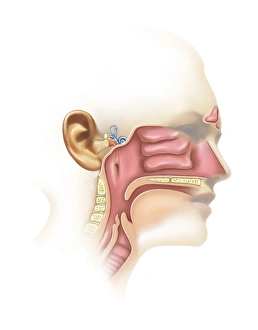
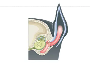
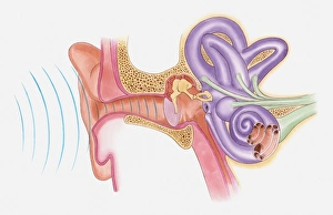
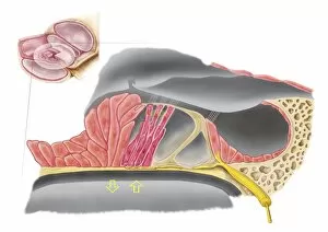
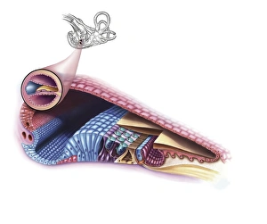
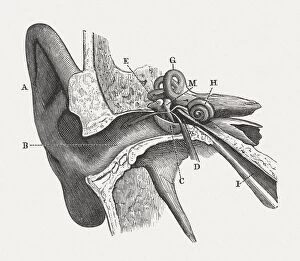
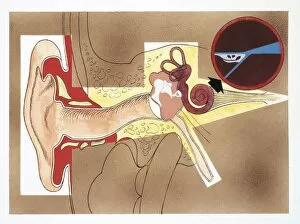
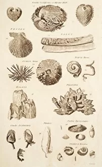
The cochlea, an intricate structure nestled within the human ear, has captivated scientists and artists alike throughout history. Dating back to 1876, a lithograph showcasing the anatomy of the human ear provides a glimpse into this remarkable organ. Adjacent to it, a diagram from the same era illustrates not only the auditory canal and eardrum but also highlights the semicircular canals and cochlea nerve. Delving further into scientific illustrations of our auditory system, we encounter captivating artwork from centuries past. A watercolor and gouache masterpiece created between 1575-80 showcases a snail with meticulous detail – perhaps hinting at its connection to the cochlea's spiral shape. Another painting features shellfish alongside marine creatures, reminding us of nature's inspiration in understanding our own biology. An engraving dedicated solely to exploring "the ear" emphasizes its significance as one of our primary senses. Meanwhile, an anatomical depiction published in 1861 sheds light on both the eye and ear's intricacies – highlighting their interdependence for perceiving sound and sight. Yet amidst these artistic interpretations lies an undeniable fascination with unraveling the mysteries hidden within our bodies' inner workings. An illustration specifically focused on dissecting the cochlear duct grants us insight into this delicate structure responsible for converting sound vibrations into electrical signals that reach our brain. Finally, we are presented with an image revealing another aspect of human hearing: The external auditory canal labeled meticulously for educational purposes. This visual aid serves as a reminder that while art may capture beauty or evoke emotions surrounding subjects like cochlea; science ultimately seeks knowledge through observation and exploration. In essence, these diverse depictions spanning centuries remind us of humanity's ceaseless curiosity about ourselves - how we perceive sound through this extraordinary organ called "cochlea.