Home > Popular Themes > Human Body
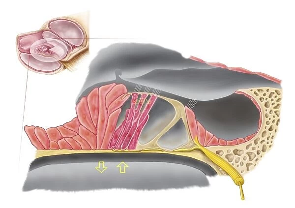
Anatomy of the organ of Corti, part of the cochlea of the inner ear
![]()

Wall Art and Photo Gifts from Stocktrek
Anatomy of the organ of Corti, part of the cochlea of the inner ear
Stocktrek Images specializes in Astronomy, Dinosaurs, Medical, Military Forces, Ocean Life, & Sci-Fi
Media ID 13013401
© Stocktrek Images
Acoustic Afferent Neurons Anatomy Audio Auditory Auditory System Aural Auricle Basilar Membrane Biology Biomedical Illustrations Cochlea Cochlear Duct Cross Section Cutaway View Detail Diagram Dissection Ear Canal Endolymph Healthcare Hearing Human Anatomy Human Body Parts Human Ear Human Organs Human Tissue Inner Ear Internal Organs Ligament Listening Magnification Medical Medicine Membrane Nerve Nerve Fibers Organ Organ Of Corti Physiology Scala Tympani Scala Vestibuli Sensory Receptors Sensory System Sound Spiral Ganglion Structure Text Vestibular Nerves Vestibular System Western Script Hair Cells Stereocilia
FEATURES IN THESE COLLECTIONS
EDITORS COMMENTS
This and intricate print showcases the mesmerizing "Anatomy of the organ of Corti, part of the cochlea of the inner ear". With its vibrant colors and digitally generated illustration, this artwork brings to life the complex world within our ears. Set against a clean white background, this medical masterpiece delves into the depths of human physiology. From ligaments to sensory receptors, every detail is meticulously depicted in this cross-section view. The cochlear duct, scala tympani, and scala vestibuli are all highlighted with precision. As we explore deeper into this auditory wonderland, we encounter essential components such as hair cells and stereocilia that play a vital role in our ability to hear. Afferent neurons and vestibular nerves connect us to our surroundings through sound waves. The artist's skillful magnification allows us to appreciate the delicate structures like tectorial membrane, Deiters cells, and Claudius cells that contribute to our auditory system's functionality. This image not only serves as an educational tool for those studying medicine or biology but also sparks curiosity about how our internal organs work harmoniously together. It reminds us of the remarkable complexity hidden within even seemingly simple bodily functions like listening. In summary, Stocktrek Images has beautifully captured an awe-inspiring representation of human anatomy with their detailed illustration depicting "Anatomy of the organ of Corti". This visually striking print invites viewers on a journey through one aspect of our incredible physiological makeup – reminding us just how extraordinary our bodies truly are.
MADE IN THE USA
Safe Shipping with 30 Day Money Back Guarantee
FREE PERSONALISATION*
We are proud to offer a range of customisation features including Personalised Captions, Color Filters and Picture Zoom Tools
SECURE PAYMENTS
We happily accept a wide range of payment options so you can pay for the things you need in the way that is most convenient for you
* Options may vary by product and licensing agreement. Zoomed Pictures can be adjusted in the Cart.

