Fraud Blocker Skip to main content

Molecular Structure Collection (page 22)

"Molecular Structure: Unlocking the Secrets of Life's Building Blocks" From anaesthetics inhibiting ion channels to antidepressant molecules

Background imageMolecular Structure Collection: Insect antifreeze protein F006 / 9319

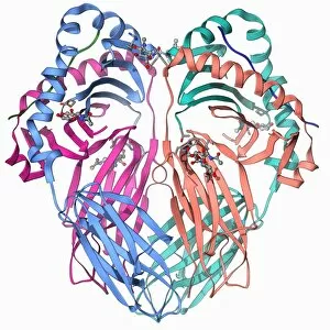
Insect antifreeze protein F006 / 9319
Insect antifreeze protein. Molecular model of an insect antifreeze protein (AFP) from the spruce budworm (Choristoneura fumiferana)

Background imageMolecular Structure Collection: Oxoguanine glycosylase complex F006 / 9318

Oxoguanine glycosylase complex F006 / 9318
Oxoguanine glycosylase complex. Computer model showing an 8-Oxoguanine glycosylase (OGG1) molecule (beige) bound to a section of DNA (deoxyribonucleic acid, red and blue)

Background imageMolecular Structure Collection: Transcription repressor molecule F006 / 9316

Transcription repressor molecule F006 / 9316
Transcription repressor. Molecular model of the Tup 1 transcription repressor protein. Transcription repressors bind to specific sequences of DNA (deoxyribonucleic acid)

Background imageMolecular Structure Collection: Restriction enzyme and DNA F006 / 9315

Restriction enzyme and DNA F006 / 9315
Restriction enzyme and DNA. Molecular model showing an EcoRI endonuclease enzyme (purple and green) bound to a DNA (deoxyribonucleic acid) molecule (red and blue)

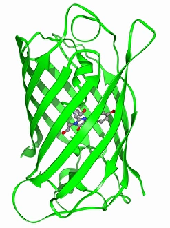
Background imageMolecular Structure Collection: Green fluorescent protein molecule F006 / 9313

Green fluorescent protein molecule F006 / 9313
Green fluorescent protein (GFP), molecular model. The molecule has a cylindrical structure formed from beta sheets (ribbons). GFP is found in the Pacific jellyfish Aequorea victoria

Background imageMolecular Structure Collection: MoaC protein molecule F006 / 9312

MoaC protein molecule F006 / 9312
MoaC protein. Molecular model of the molybdenum cofactor biosynthesis protein MoaC. This enzyme is involved in carbon, nitrogen and sulphur metabolism

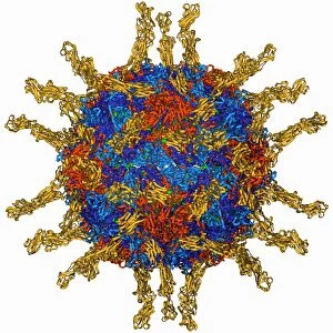
Background imageMolecular Structure Collection: Poliovirus particle F006 / 9306

Poliovirus particle F006 / 9306
Poliovirus particle, molecular model

Background imageMolecular Structure Collection: LAC repressor bound to DNA F006 / 9309

LAC repressor bound to DNA F006 / 9309
LAC repressor bound to DNA. Molecular model of a LAC (lactose) repressor molecule (pink and turquoise) interacting with bacterial DNA (deoxyribonucleic acid, red and blue)

Background imageMolecular Structure Collection: Epidermal growth factor molecule F006 / 9311

Epidermal growth factor molecule F006 / 9311
Epidermal growth factor (EGF), molecular model. EGF plays an important role in the regulation of cell growth, proliferation and differentiation

Background imageMolecular Structure Collection: Elongation factors Tu and Ts F006 / 9310

Elongation factors Tu and Ts F006 / 9310
Elongation factors Tu and Ts, molecular model. These enzymes are involved in the elongation of polypeptide chains during translation

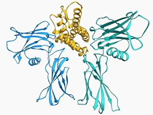
Background imageMolecular Structure Collection: Erythropoietin bound to receptors F006 / 9308

Erythropoietin bound to receptors F006 / 9308
Artwork of the human erythropoietin (EPO) hormone molecule (yellow) bound to receptors (pink). Erythropoietin regulates blood oxygen levels in the body

Background imageMolecular Structure Collection: Pyruvate dehydrogenase complex enzyme F006 / 9303

Pyruvate dehydrogenase complex enzyme F006 / 9303
Pyruvate dehydrogenase complex enzyme. Molecular model of dihydrolipoyl transacetylase one of the enzymes in the pyruvate dehydrogenase complex

Background imageMolecular Structure Collection: Oxoguanine glycosylase complex F006 / 9307

Oxoguanine glycosylase complex F006 / 9307
Oxoguanine glycosylase complex. Computer model showing an 8-Oxoguanine glycosylase (OGG1) molecule (beige) bound to a section of DNA (deoxyribonucleic acid, red and blue)

Background imageMolecular Structure Collection: Semliki forest virus capsid F006 / 9297

Semliki forest virus capsid F006 / 9297
Semliki forest virus capsid, molecular model. This virus, named for the forest in Uganda where it was identified, is spread by the bite of mosquitoes. It can infect both humans and animals

Background imageMolecular Structure Collection: ATPase molecule F006 / 9300

ATPase molecule F006 / 9300
ATPase molecule. Molecular model of the central stalk of an ATP synthase (ATPase) molecule from a cow. ATPase is an important enzyme that provides energy for cells through the synthesis of adenosine

Background imageMolecular Structure Collection: Cytochrome P450 and fluconazole F006 / 9302

Cytochrome P450 and fluconazole F006 / 9302
Cytochrome P450 and fluconazole. Molecular model of cytochrome P450 complexed with the anti-fungal compound fluconazole. This protein plays a crucial role in metabolism in animals (including humans)

Background imageMolecular Structure Collection: Bacteriorhodopsin protein F006 / 9299

Bacteriorhodopsin protein F006 / 9299
Bacteriorhodopsin protein. Molecular model showing the structure of bacteriorhodopsin (bR), a protein found in primitive micro-organisms known as Archaea. This protein acts as a proton pump

Background imageMolecular Structure Collection: Human serum albumin molecule F006 / 9301

Human serum albumin molecule F006 / 9301
Human serum albumin, molecular model. Albumin is the most abundant protein in human blood plasma. It is seen here complexed with stearic acid, a saturated fatty acid

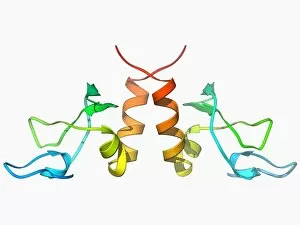
Background imageMolecular Structure Collection: HP1 molecule C-terminal domain F006 / 9298

HP1 molecule C-terminal domain F006 / 9298
HP1 molecule C-terminal domain. Molecular model showing the structure of the C terminal (shadow chromo) domain of a heterochromatin protein 1 (HP1) molecule from a mouse

Background imageMolecular Structure Collection: Human poliovirus, molecular model F006 / 9289

Human poliovirus, molecular model F006 / 9289
Human poliovirus particle. Molecular model of the capsid of the human poliovirus. The capsid is a protein coat that encloses the viruss genetic information (genome), stored as RNA (ribonucleic acid)

Background imageMolecular Structure Collection: Green mamba venom molecule F006 / 9296

Green mamba venom molecule F006 / 9296
Green mamba venom. Molecular model of the alpha-dendrotoxin from green mamba (Dendroaspis angusticeps) venom

Background imageMolecular Structure Collection: MHC protein complexed with flu virus F006 / 9294

MHC protein complexed with flu virus F006 / 9294
MHC protein complexed with flu virus. Molecular model showing human class II MHC (major histocompatibility complex) protein HLA-DR1 complexed with an influenza (flu) virus peptide

Background imageMolecular Structure Collection: Hepatitis D virus ribozyme complex F006 / 9295

Hepatitis D virus ribozyme complex F006 / 9295
Hepatitis D virus ribozyme complex. Molecular model showing an RNA (ribonucleic acid) strand from an Hepatitis delta (Hepatitis D) virus genomic ribozyme, complexed with a ribonucleoprotein

Background imageMolecular Structure Collection: Heat shock protein 70 F006 / 9293

Heat shock protein 70 F006 / 9293
Heat shock protein 70. Molecular model of the 70 kDa heat shock protein (HSP) complexed with its substrate peptide. HSPs are a group of proteins whose levels increase when cells are exposed to raised

Background imageMolecular Structure Collection: Nucleotide exchange factor F006 / 9292

Nucleotide exchange factor F006 / 9292
Nucleotide exchange factor. Molecular model of the nucleotide exchange factor protein GrpE complexed with the chaperone protein DnaK

Background imageMolecular Structure Collection: Lumazine synthase molecule F006 / 9291

Lumazine synthase molecule F006 / 9291
Lumazine synthase molecule. Molecular model showing the structure of a lumazine synthase enzyme molecule from a Brucella abortus bacterium

Background imageMolecular Structure Collection: NAD-dependent DNA ligase molecule F006 / 9290

NAD-dependent DNA ligase molecule F006 / 9290
NAD dependent DNA ligase. Molecular model of NAD(+)-dependent DNA ligase. DNA ligase is an enzyme that binds two strands of DNA (deoxyribonucleic acid) together

Background imageMolecular Structure Collection: Human catalsae, molecular model F006 / 9288

Human catalsae, molecular model F006 / 9288
Human catalase, molecular model. This enzyme catalyses the break down of hydrogen peroxide to water and oxygen. Hydrogen peroxide is a highly toxic byproduct of a number of normal cellular processes

Background imageMolecular Structure Collection: Beta-carbonic anhydrase molecule F006 / 9286

Beta-carbonic anhydrase molecule F006 / 9286
Beta-carbonic anhydrase, molecular model. This enzyme is from the red algae Porphyridium purpureum. It catalyses the reversible hydration of carbon dioxide

Background imageMolecular Structure Collection: Ribonuclease bound to inhibitor F006 / 9287

Ribonuclease bound to inhibitor F006 / 9287
Ribonuclease bound to inhibitor, molecular model. Ribonuclease (RNase) is a type of nuclease that catalyses the degradation of RNA (ribonucleic acid)

Background imageMolecular Structure Collection: Elongation factor G F006 / 9284

Elongation factor G F006 / 9284
Elongation factor G. Molecular model of elongation factor G (EF-G) complexed with GDP (guanosine diphosphate). This enzyme is involved in the elongation of polypeptide chains during translation

Background imageMolecular Structure Collection: DNA Holliday junction, molecular model F006 / 9285

DNA Holliday junction, molecular model F006 / 9285
DNA Holliday junction. Molecular model of a Holliday junction (centre) between homologous strands of DNA (deoxyribonucleic acid)

Background imageMolecular Structure Collection: Coagulation factor complex molecule F006 / 9283

Coagulation factor complex molecule F006 / 9283
Coagulation factor complex molecule. Molecular model showing a blood clotting factor VIIa-tissue factor complex molecule. Tissue factor, also known as factor III

Background imageMolecular Structure Collection: Yeast DNA recognition, molecular model F006 / 9282

Yeast DNA recognition, molecular model F006 / 9282
Yeast DNA recognition. Computer model showing a GAL4 transcription activator protein bound to a yeast DNA (deoxyribonucleic acid) molecule (red and blue)

Background imageMolecular Structure Collection: Creatine kinase molecule F006 / 9276

Creatine kinase molecule F006 / 9276
Creatine kinase molecule. This enzyme catalyses the phosphorylation of creatine to creatine phosphate. It plays an important role in energy metabolism in cells with high or fluctuating energy

Background imageMolecular Structure Collection: Human transferrin receptor molecule F006 / 9280

Human transferrin receptor molecule F006 / 9280
Human transferrin receptor, molecular model. This molecule is found on the surface of a cell. It binds transferrins, iron-binding glycoproteins found in the blood plasma

Background imageMolecular Structure Collection: Sex hormone-binding globulin molecule F006 / 9281

Sex hormone-binding globulin molecule F006 / 9281
Sex hormone-binding globulin. Molecular model of the sex hormone-binding globulin (SHBG) protein complexed with the male sex hormone dihydrotestosterone

Background imageMolecular Structure Collection: Adenylyl cyclase enzyme molecule F006 / 9279

Adenylyl cyclase enzyme molecule F006 / 9279
Adenylyl cyclase. Molecular model of adenylyl cyclase complexed with an inhibitor. This enzyme catalyses the conversion of ATP (adenosine triphosphate) to cyclic AMP (cAMP) and pyrophosphate

Background imageMolecular Structure Collection: Citrate synthase molecule F006 / 9277

Citrate synthase molecule F006 / 9277
Citrate synthase, molecular model. This enzyme is involved in the first step of the citric acid (or Krebs) cycle, the process by which mitochondria convert glucose to energy

Background imageMolecular Structure Collection: Cobra venom molecule F006 / 9278

Cobra venom molecule F006 / 9278
Cobra venom. Molecular model of an alpha-cobratoxin (snake venom protein) from an Indochinese spitting cobra (Naja siamensis)

Background imageMolecular Structure Collection: DNA clamp complexed with DNA molecule F006 / 9274

DNA clamp complexed with DNA molecule F006 / 9274
DNA clamp complexed with DNA molecule. Molecular model showing a sliding DNA (deoxyribonucleic acid) clamp (beige) complexed with a molecule of DNA (red and blue)

Background imageMolecular Structure Collection: Synthetic triple helical peptide molecule F006 / 9275

Synthetic triple helical peptide molecule F006 / 9275
Synthetic triple helical peptide, molecular model

Background imageMolecular Structure Collection: mRNA capping enzyme molecule F006 / 9273

mRNA capping enzyme molecule F006 / 9273
mRNA capping enzyme. Molecular model of the mRNA capping enzyme mRNA guanylyltransferase complexed with a cap analogue

Background imageMolecular Structure Collection: Adenylyl cyclase enzyme molecule F006 / 9271

Adenylyl cyclase enzyme molecule F006 / 9271
Adenylyl cyclase enzyme, molecular model. This enzyme catalyses the conversion of ATP (adenosine triphosphate) to cyclic AMP (cAMP) and pyrophosphate. cAMP regulates numerous cell functions

Background imageMolecular Structure Collection: Serotonin N-acetyl transferase molecule F006 / 9272

Serotonin N-acetyl transferase molecule F006 / 9272
Serotonin N-acetyl transferase. Molecular model of serotonin N-acetyl transferase complexed with coenzyme A-S-acetyltryptamine

Background imageMolecular Structure Collection: HGPRTase molecule F006 / 9270

HGPRTase molecule F006 / 9270
HGPRTase. Molecular model of hypoxanthine-guanine phosphoribosyltransferase (HGPRTase) from the malaria parasite Plasmodium falciparum. HGPRTase is involved in the purine salvage pathway

Background imageMolecular Structure Collection: Bleomycin hydrolase molecule F006 / 9265

Bleomycin hydrolase molecule F006 / 9265
Bleomycin hydrolase. Molecular model of the cysteine protease bleomycin hydrolase. This enzyme is thought to be a major cause of resistance to the bleomycin chemotherapy drug

Background imageMolecular Structure Collection: Gene activator protein F006 / 9269

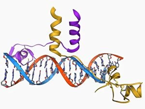
Gene activator protein F006 / 9269
Gene activator protein. Molecular model of catabolite gene activator protein (CAP, pink and green) bound to a molecule of deoxyribonucleic acid (DNA, across top)


All Professionally Made to Order for Quick Shipping

"Molecular Structure: Unlocking the Secrets of Life's Building Blocks" From anaesthetics inhibiting ion channels to antidepressant molecules, the intricate world holds endless wonders. The C015 / 6718 anaesthetic molecule delicately interacts with ion channels, altering their function and providing relief from pain. Meanwhile, Amitriptyline, an antidepressant molecule, works its magic by modulating neurotransmitters in our brains. In the realm of immunity, Immunoglobulin G antibody F007 / 9894 stands tall as a defender against pathogens. Its unique structure allows it to recognize and neutralize foreign invaders effectively. On another front, DNA artwork showcases the elegance and complexity that underlies all life forms on Earth. Creatine amino acid molecule fuels our muscles during intense physical activities while nanotube technology revolutionizes various industries with its exceptional properties. These tiny tubes hold immense potential for advancements in medicine and materials science alike. Zinc fingers bound to a DNA strand demonstrate how proteins can precisely interact with genetic material. This interaction plays a crucial role in gene regulation and expression. Carbon nanotubes take center stage once again as they exhibit remarkable strength and conductivity at the nano-scale level. Oxytocin neurotransmitter molecule reminds us of love's powerful influence on human connections—its presence promotes bonding between individuals. Manganese superoxide dismutase enzyme F006 / 9423 safeguards our cells by combating harmful free radicals that contribute to aging and disease. Even viruses have their own molecular structures; SARS coronavirus protein represents one such example—a key player in viral replication within host cells. Conceptual artwork further explores nanotube technology's limitless possibilities—the fusion of imagination and scientific innovation knows no bounds here. As we delve deeper into understanding molecular structures, we unravel nature's blueprint for life itself—one atom at a time. These captivating glimpses into the microscopic world remind us of both the fragility and resilience found within the building blocks of existence.