Fraud Blocker Skip to main content

Molecular Structure Collection (page 19)

"Molecular Structure: Unlocking the Secrets of Life's Building Blocks" From anaesthetics inhibiting ion channels to antidepressant molecules

Background imageMolecular Structure Collection: Aspirin drug target molecule F006 / 9464

Aspirin drug target molecule F006 / 9464
Aspirin drug target. Molecular model of the enzyme prostaglandin H2 synthase (PGHS), the target of the anti-inflammatory drug aspirin

Background imageMolecular Structure Collection: Archaeon enzyme, molecular model F006 / 9459

Archaeon enzyme, molecular model F006 / 9459
Archaeon enzyme. Molecular model of an enzyme from Thermoplasma acidophilum. This is the 20S proteasome. A proteasome is a complex type of proteinase (protein-digesting enzyme)
Background imageMolecular Structure Collection: Whooping cough toxin molecule F006 / 9463

Whooping cough toxin molecule F006 / 9463
Whooping cough toxin. Molecular model of the pertussis toxin from the bacterium Bordetella pertussis, the cause of whooping cough

Background imageMolecular Structure Collection: Photosynthetic reaction centre F006 / 9462

Photosynthetic reaction centre F006 / 9462
Photosynthetic reaction centre. Molecular model of the photosynthetic reaction centre from the purple bacterium Rhodopseudomonas viridis

Background imageMolecular Structure Collection: Dengue virus capsid, molecular model F006 / 9455

Dengue virus capsid, molecular model F006 / 9455
Dengue virus capsid, molecular model. This virus, transmitted by mosquito bites, causes the tropical disease dengue fever in humans

Background imageMolecular Structure Collection: Amylase, molecular model F006 / 9461

Amylase, molecular model F006 / 9461
Amylase. Molecular model of the enzyme alpha-amylase from the human pancreas. Amylase catalyses the breakdown of starch to sugars, and so starts the digestive process

Background imageMolecular Structure Collection: GST enzyme conferring DDT resistance F006 / 9460

GST enzyme conferring DDT resistance F006 / 9460
GST enzyme. Molecular model of a glutathione S-transferase (GST) enzyme from the malaria-carrying mosquito Anopheles gambiae

Background imageMolecular Structure Collection: Caspase-3 and inhibitor F006 / 9457

Caspase-3 and inhibitor F006 / 9457
Apopain protein and inhibitor. Molecular model of caspase-3, also known as apopain, complexed with an inhibitor. Caspase-3 is a protease, an enzyme that cleaves proteins

Background imageMolecular Structure Collection: Para-hydroxybenzoate hydroxylase molecule F006 / 9458

Para-hydroxybenzoate hydroxylase molecule F006 / 9458
Para-hydroxybenzoate hydroxylase, molecular model. This flavoprotein enzyme is involved in the degradation of aromatic compounds

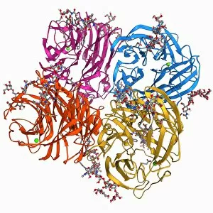
Background imageMolecular Structure Collection: Adenovirus hexon protein F006 / 9453

Adenovirus hexon protein F006 / 9453
Adenovirus hexon protein, molecular model. Hexon proteins are part of the protein coat or shell (capsid) of adenoviruses. In viruses

Background imageMolecular Structure Collection: Rous sarcoma virus capsid protein F006 / 9456

Rous sarcoma virus capsid protein F006 / 9456
Rous sarcoma virus capsid protein. Molecular model of the N-terminal domain of the Rous sarcoma virus capsid protein

Background imageMolecular Structure Collection: Nitric oxide synthase molecule F006 / 9452

Nitric oxide synthase molecule F006 / 9452
Nitric oxide synthase, molecular model. This enzyme catalyses the production of nitric oxide from L-arginine. Nitric oxide is involved in cellular signalling

Background imageMolecular Structure Collection: P32 mitochondrial matrix protein F006 / 9454

P32 mitochondrial matrix protein F006 / 9454
P32 mitochondrial matrix protein, molecular model. Also known as SF2-associated p32 (SF2P32), this protein is found in the matrix of cellular mitochondria

Background imageMolecular Structure Collection: Cytochrome c oxidase molecule F006 / 9447

Cytochrome c oxidase molecule F006 / 9447
Cytochrome c oxidase. Molecular model of a cytochrome c oxidase enzyme from the mitochondria of a cows heart. Cytochrome molecules perform oxidation and reduction reactions for electron transport

Background imageMolecular Structure Collection: ATPase and inhibitor, molecular model F006 / 9448

ATPase and inhibitor, molecular model F006 / 9448
ATPase and inhibitor. Computer model of an ATP synthase (ATPase) molecule from a mitochondrion complexed with its inhibitor protein IF1

Background imageMolecular Structure Collection: Dengue virus surface protein molecule F006 / 9449

Dengue virus surface protein molecule F006 / 9449
Dengue virus surface protein molecule. Molecular model of the envelope glycoprotein found on the surface of the dengue haemorrhagic fever (DHF) virus

Background imageMolecular Structure Collection: Cytochrome c oxidase molecule F006 / 9446

Cytochrome c oxidase molecule F006 / 9446
Cytochrome c oxidase. Molecular model of a cytochrome c oxidase enzyme from the mitochondria of a cows heart. Cytochrome molecules perform oxidation and reduction reactions for electron transport

Background imageMolecular Structure Collection: Tumour suppressor protein molecular model F006 / 9450

Tumour suppressor protein molecular model F006 / 9450
Tumour suppressor protein, molecular model. P53 prevents the proliferation of cells with damaged DNA. Its production occurs in response to radiation and chemicals that damage the structure of DNA

Background imageMolecular Structure Collection: Shrew eye lens protein molecule F006 / 9444

Shrew eye lens protein molecule F006 / 9444
Shrew eye lens protein. Molecular model of eta-crystallin, a structural protein only found in the eye lenses of elephant shrews (family Macroscelididae)

Background imageMolecular Structure Collection: Nodamura virus capsid, molecular model F006 / 9438

Nodamura virus capsid, molecular model F006 / 9438
Nodamura virus capsid, molecular model. This virus can cause lethal infections in both mammals and insects. In viruses, the capsid is the protein shell that encloses the genetic material

Background imageMolecular Structure Collection: Retinoblastoma tumor suppressor molecule F006 / 9445

Retinoblastoma tumor suppressor molecule F006 / 9445
Retinoblastoma tumour suppressor. Molecular model of a retinoblastoma tumour suppressor (Rb) bound to a transcription factor E2F-1

Background imageMolecular Structure Collection: Citrate synthase molecule F006 / 9443

Citrate synthase molecule F006 / 9443
Citrate synthase, molecular model. This enzyme is involved in the first step of the citric acid (or Krebs) cycle, the process by which mitochondria convert glucose to energy

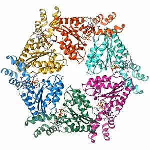
Background imageMolecular Structure Collection: ATPase molecule F006 / 9441

ATPase molecule F006 / 9441
ATPase molecule. Molecular model of the ATP-dependent oligomerisation domain of N-ethylmaleimide sensitive factor (NSF), a hexameric ATPase

Background imageMolecular Structure Collection: Caspase-9 with inhibitor, molecular model F006 / 9442

Caspase-9 with inhibitor, molecular model F006 / 9442
Caspase-9 complexed with an inhibitor, molecular model. Caspase-9 is a protease, an enzyme that cleaves proteins, that plays a role in apoptosis (programmed cell death)

Background imageMolecular Structure Collection: Epidermal growth factor and receptor F006 / 9440

Epidermal growth factor and receptor F006 / 9440
Epidermal growth factor molecule. Molecular model of epidermal growth factor (EGF) bound to a receptor. EGF plays an important role in the regulation of cell growth, proliferation and differentiation

Background imageMolecular Structure Collection: Oxytocin and carrier protein F006 / 9439

Oxytocin and carrier protein F006 / 9439
Oxytocin and carrier protein. Molecular model of the hormone oxytocin bound to its carrier protein neurophysin I. Oxytocin is a neurotransmitter that plays a role in labour, sexual arousal, bonding

Background imageMolecular Structure Collection: Cytochrome P450 F006 / 9437

Cytochrome P450 F006 / 9437
Cytochrome P450. Molecular model of cytochrome P450 complexed with its catalytic product hydroxycamphor. This protein plays a crucial role in metabolism in animals (including humans), fungi

Background imageMolecular Structure Collection: DNA transcription, molecular model F006 / 9424

DNA transcription, molecular model F006 / 9424
DNA transcription. Molecular model of the enzyme RNA polymerase II synthesising a mRNA (messenger ribonucleic acid) strand from a DNA (deoxyribonucleic acid) template

Background imageMolecular Structure Collection: Nitric oxide synthase molecule F006 / 9436

Nitric oxide synthase molecule F006 / 9436
Nitric oxide synthase, molecular model. This enzyme catalyses the production of nitric oxide from L-arginine. Nitric oxide is involved in cellular signalling

Background imageMolecular Structure Collection: Human poliovirus, molecular model F006 / 9434

Human poliovirus, molecular model F006 / 9434
Human poliovirus particle. Molecular model of the capsid of the human poliovirus. The capsid is a protein coat that encloses the viruss genetic information (genome), stored as RNA (ribonucleic acid)

Background imageMolecular Structure Collection: VEE equine encephalitis virus capsid

VEE equine encephalitis virus capsid
Venezuelan equine encephalitis virus capsid, molecular model. This mosquito-borne virus can kill horses and other equine species, causing brain and spinal cord inflammation

Background imageMolecular Structure Collection: Herpesvirus capsid protein F006 / 9435

Herpesvirus capsid protein F006 / 9435
Herpesvirus capsid protein. Molecular model of the major capsid protein VP5 from herpes simplex virus-1 (HSV-1). This virus infects epithelial cells around the mouth

Background imageMolecular Structure Collection: Rhinovirus 14 capsid, molecular model F006 / 9430

Rhinovirus 14 capsid, molecular model F006 / 9430
Rhinovirus 14 capsid, molecular model. This is human rhinovirus 14. The rhinovirus infects the upper respiratory tract and is the cause of the common cold. It is spread by coughs and sneezes

Background imageMolecular Structure Collection: Flu virus surface protein molecule F006 / 9433

Flu virus surface protein molecule F006 / 9433
Flu virus surface protein molecule. Molecular model of the neuraminidase glycoprotein enzyme found on the surface of the influenza A (flu) virus

Background imageMolecular Structure Collection: Succinate dehydrogenase enzyme F006 / 9432

Succinate dehydrogenase enzyme F006 / 9432
Succinate dehydrogenase enzyme. Molecular model of the succinate dehydrogenase (complex II) enzyme from an Escherichia coli bacterium

Background imageMolecular Structure Collection: Flu virus surface protein and antibody F006 / 9428

Flu virus surface protein and antibody F006 / 9428
Flu virus surface protein and antibody. Molecular model of the neuraminidase glycoprotein enzyme found on the surface of the influenza (flu) virus

Background imageMolecular Structure Collection: Cell adhesion protein molecule F006 / 9429

Cell adhesion protein molecule F006 / 9429
Cell adhesion protein. Molecular model of the cell adhesion protein n-cadherin (neural cadherin). This transmembrane protein facilitates adhesion between cells

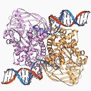
Background imageMolecular Structure Collection: Transposase enzyme and DNA complex F006 / 9425

Transposase enzyme and DNA complex F006 / 9425
Transposase enzyme and DNA complex. Molecular model of a TN5 transposase enzyme bound to the transposon end of a DNA molecule (red and blue)

Background imageMolecular Structure Collection: DNA helicase molecule F006 / 9426

DNA helicase molecule F006 / 9426
DNA helicase. Molecular model of a helicase molecule from the SV40 virus. Helicases are enzymes that separate the two strands of the DNA double helix

Background imageMolecular Structure Collection: Hepatitis C polymerase enzyme F006 / 9427

Hepatitis C polymerase enzyme F006 / 9427
Hepatitis C polymerase enzyme, molecular model. This protein is the NS5b RNA polymerase found in the virus hepatitis C (genotype-1b, strain J4)

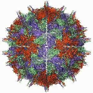
Background imageMolecular Structure Collection: Bacteriophage alpha 3 capsid F006 / 9414

Bacteriophage alpha 3 capsid F006 / 9414
Bacteriophage alpha 3 capsid, molecular model. Bacteriophages are viruses that infect bacteria. Bacteriophage alpha 3 has a capsid consisting of 60 coat proteins (blue) and 12 pentameric (5-subunit)

Background imageMolecular Structure Collection: Hammerhead ribozyme molecule F006 / 9422

Hammerhead ribozyme molecule F006 / 9422
Hammerhead ribozyme, molecular model. Ribozymes are RNA (ribonucleic acid) molecules that catalyse certain biochemical reactions

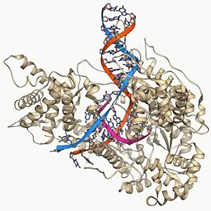
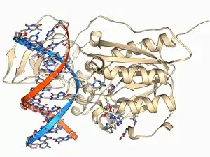
Background imageMolecular Structure Collection: Methyltransferase and DNA F006 / 9421

Methyltransferase and DNA F006 / 9421
Methyltransferase and DNA. Molecular model of the enzyme HhaI methyltransferase (beige) complexed with a molecule of DNA (deoxyribonucleic acid, red and blue)

Background imageMolecular Structure Collection: Insect flight muscle, molecular model F006 / 9416

Insect flight muscle, molecular model F006 / 9416
Insect flight muscle. Molecular model of myosin crossbridges in insect flight muscles in the nucleotide-free (rigor) state. This conformation is thought to represent the end of the power stroke

Background imageMolecular Structure Collection: Diphtheria toxin, molecular model F006 / 9419

Diphtheria toxin, molecular model F006 / 9419
Diphtheria toxin, molecular model. Diphtheria is caused by the bacterium Corynebacterium diphtheriae. Symptoms include sore throat, fever and breathing difficulties

Background imageMolecular Structure Collection: Myoglobin molecule F006 / 9418

Myoglobin molecule F006 / 9418
Myoglobin, molecular model. Myoglobin is a protein found in muscle tissue, where it binds to and stores oxygen to be used during strenuous exercise

Background imageMolecular Structure Collection: Myoglobin molecule F006 / 9417

Myoglobin molecule F006 / 9417
Myoglobin, molecular model. Myoglobin is a protein found in muscle tissue, where it binds to and stores oxygen to be used during strenuous exercise

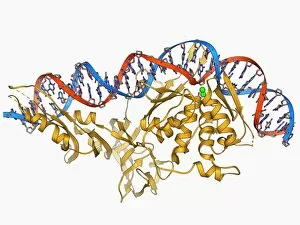
Background imageMolecular Structure Collection: Endonuclease and DNA, molecular model F006 / 9413

Endonuclease and DNA, molecular model F006 / 9413
Endonuclease and DNA. Molecular model of an endonuclease restriction enzyme (yellow) bound to a molecule of DNA (deoxyribonucleic acid)


All Professionally Made to Order for Quick Shipping

"Molecular Structure: Unlocking the Secrets of Life's Building Blocks" From anaesthetics inhibiting ion channels to antidepressant molecules, the intricate world holds endless wonders. The C015 / 6718 anaesthetic molecule delicately interacts with ion channels, altering their function and providing relief from pain. Meanwhile, Amitriptyline, an antidepressant molecule, works its magic by modulating neurotransmitters in our brains. In the realm of immunity, Immunoglobulin G antibody F007 / 9894 stands tall as a defender against pathogens. Its unique structure allows it to recognize and neutralize foreign invaders effectively. On another front, DNA artwork showcases the elegance and complexity that underlies all life forms on Earth. Creatine amino acid molecule fuels our muscles during intense physical activities while nanotube technology revolutionizes various industries with its exceptional properties. These tiny tubes hold immense potential for advancements in medicine and materials science alike. Zinc fingers bound to a DNA strand demonstrate how proteins can precisely interact with genetic material. This interaction plays a crucial role in gene regulation and expression. Carbon nanotubes take center stage once again as they exhibit remarkable strength and conductivity at the nano-scale level. Oxytocin neurotransmitter molecule reminds us of love's powerful influence on human connections—its presence promotes bonding between individuals. Manganese superoxide dismutase enzyme F006 / 9423 safeguards our cells by combating harmful free radicals that contribute to aging and disease. Even viruses have their own molecular structures; SARS coronavirus protein represents one such example—a key player in viral replication within host cells. Conceptual artwork further explores nanotube technology's limitless possibilities—the fusion of imagination and scientific innovation knows no bounds here. As we delve deeper into understanding molecular structures, we unravel nature's blueprint for life itself—one atom at a time. These captivating glimpses into the microscopic world remind us of both the fragility and resilience found within the building blocks of existence.