Fraud Blocker Skip to main content

Molecular Structure Collection (page 13)

"Molecular Structure: Unlocking the Secrets of Life's Building Blocks" From anaesthetics inhibiting ion channels to antidepressant molecules

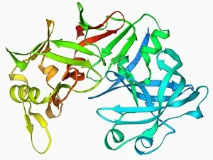
Background imageMolecular Structure Collection: Pepsin stomach enzyme F006 / 9767

Pepsin stomach enzyme F006 / 9767
Pepsin stomach enzyme, molecular model. Pepsin is a protease enzyme that is secreted as part of gastric juice into the stomach in an inactive form known as pepsinogen

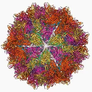
Background imageMolecular Structure Collection: Flock house virus capsid F006 / 9755

Flock house virus capsid F006 / 9755
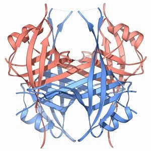
Flock house virus capsid, molecular model. The flock house virus is a member of the Nodaviridae family. It kills the New Zealand grass grub insect

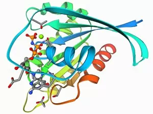
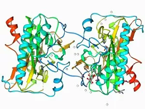
Background imageMolecular Structure Collection: Xylose isomerase complex F006 / 9765

Xylose isomerase complex F006 / 9765
Xylose isomerase complex. Molecular model of the enzyme D-xylose isomerase bound to the sugar alcohol sorbitol. D-xylose isomerase is involved in fructose and mannose metabolism

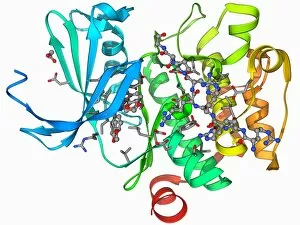
Background imageMolecular Structure Collection: H-Ras p21 oncogene protein F006 / 9766

H-Ras p21 oncogene protein F006 / 9766
H-Ras p21 oncogene protein, molecular model. The Ras proteins are involved in transmitting signals within cells. Excessive signalling can lead to conditions such as cancer

Background imageMolecular Structure Collection: Phosphofructokinase bacterial enzyme F006 / 9762

Phosphofructokinase bacterial enzyme F006 / 9762
Phosphofructokinase enzyme, molecular model. This enzyme, from the bacterium Bacillus stearothermophilus, is involved in regulating the process of releasing energy from glucose

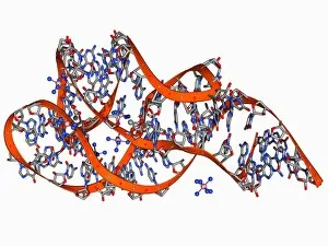
Background imageMolecular Structure Collection: tRNA molecule F006 / 9764

tRNA molecule F006 / 9764
Transfer RNA (tRNA), molecular model. tRNA (transfer ribonucleic acid) translates messenger RNA (mRNA) into a protein product

Background imageMolecular Structure Collection: H-Ras p21 oncogene protein F006 / 9763

H-Ras p21 oncogene protein F006 / 9763
H-Ras p21 oncogene protein, molecular model. The Ras proteins are involved in transmitting signals within cells. Excessive signalling can lead to conditions such as cancer

Background imageMolecular Structure Collection: Kinase inhibitor complex F006 / 9760

Kinase inhibitor complex F006 / 9760
Kinase inhibitor complex. Molecular model of a leucettine kinase inhibitor bound to a serine threonine kinase protein

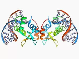
Background imageMolecular Structure Collection: Marburg viral protein 35 and RNA F006 / 9759

Marburg viral protein 35 and RNA F006 / 9759
Marburg viral protein 35 and RNA. Molecular model of the Marburg viral protein 35 (VP35) bound to a molecule of double stranded RNA (ribonucleic acid)

Background imageMolecular Structure Collection: Insulin molecule F006 / 9761

Insulin molecule F006 / 9761
Insulin molecule. Molecular model of the hormone insulin from a pig. Insulin consists of two peptide chains, A and B, which are linked by disulphide bridges
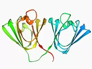
Background imageMolecular Structure Collection: Eye lens protein molecule F006 / 9758

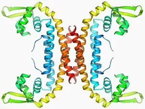
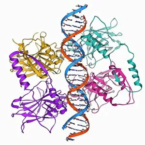
Eye lens protein molecule F006 / 9758
Eye lens protein. Molecular model of gammaB-crystallin, a protein found in the lens of the eye. The regular arrangement of the protein in the lens is thought to be responsible for its transparency

Background imageMolecular Structure Collection: Methionine aminopeptidase molecule F006 / 9756

Methionine aminopeptidase molecule F006 / 9756
Methionine aminopeptidase, molecular model. This enzyme removes the amino acid methionine from proteins

Background imageMolecular Structure Collection: Hepatitis C glycoprotein and antibody F006 / 9757

Hepatitis C glycoprotein and antibody F006 / 9757
Hepatitis C glycoprotein and antibody. Molecular model of the E2 envelope glycoprotein from the hepatitis C virus bound to a neutralising antibody

Background imageMolecular Structure Collection: Malignant brain-tumor-like protein F006 / 9754

Malignant brain-tumor-like protein F006 / 9754
Malignant brain-tumour-like protein. Molecular model of the human lethal(3) malignant brain-tumour-like protein (Malignant brain-tumour-like protein (L3MBTL3)

Background imageMolecular Structure Collection: Guanine-responsive riboswitch F006 / 9753

Guanine-responsive riboswitch F006 / 9753
Guanine-responsive riboswitch, molecular model. This protein regulates gene expression by binding to the nucleotide guanine to switch off transcription

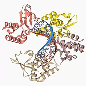
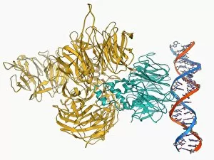
Background imageMolecular Structure Collection: UV-damaged DNA-binding protein and DNA F006 / 9750

UV-damaged DNA-binding protein and DNA F006 / 9750
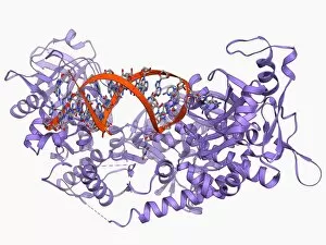
UV-damaged DNA-binding protein and DNA. Molecular model of UV-damaged DNA-binding protein (UV-DDB) complexed with DNA (deoxyribonucleic acid, red and blue)

Background imageMolecular Structure Collection: Plexin signal transduction molecule F006 / 9751

Plexin signal transduction molecule F006 / 9751
Plexin signal transduction molecule. Molecular model of plexin-A4 a signal transduction protein that is involved in neural maintenance and regeneration

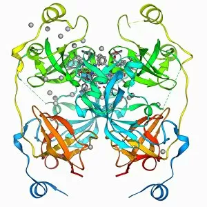
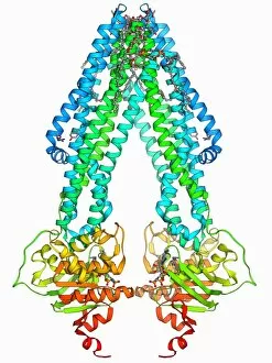
Background imageMolecular Structure Collection: Multidrug efflux pump molecule F006 / 9748

Multidrug efflux pump molecule F006 / 9748
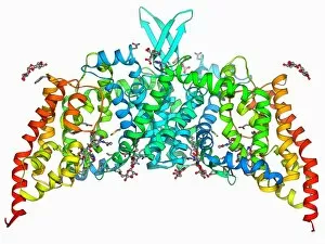
Multidrug efflux pump. Molecular model of the multidrug efflux pump AcrB from the bacterium Escherichia coli transporting two doxorubicin molecules

Background imageMolecular Structure Collection: RNA triplet repeat expansion F006 / 9749

RNA triplet repeat expansion F006 / 9749
RNA triplet repeat expansion. Molecular model of a CUG triplet repeat expansion in a molecule of double stranded RNA (ribonucleic acid)

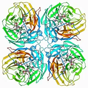
Background imageMolecular Structure Collection: Flu virus surface protein and drug F006 / 9745

Flu virus surface protein and drug F006 / 9745
Flu virus surface protein and drug. Molecular model of the neuraminidase glycoprotein enzyme from on the surface of the influenza A (flu) virus bound to the drug zanamivir

Background imageMolecular Structure Collection: Human lysine-specific demethylase F006 / 9747

Human lysine-specific demethylase F006 / 9747
Human lysine-specific demethylase, molecular model. This enzyme removes methyl groups from lysine residues in histones (proteins that package DNA)

Background imageMolecular Structure Collection: Rhinovirus capsid, molecular model F006 / 9737

Rhinovirus capsid, molecular model F006 / 9737
Rhinovirus capsid, molecular model. This is human rhinovirus. The rhinovirus infects the upper respiratory tract and is the cause of the common cold. It is spread by coughs and sneezes

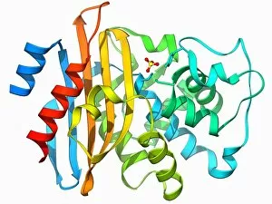
Background imageMolecular Structure Collection: Beta-lactamase molecule F006 / 9746

Beta-lactamase molecule F006 / 9746
Beta-lactamase enzyme, molecular model. This bacterial protein provides resistance to beta-lactam antibiotics, which include penicillin

Background imageMolecular Structure Collection: Human muscle aldolase, molecular model F006 / 9742

Human muscle aldolase, molecular model F006 / 9742
Human muscle aldolase. Molecular model of the enzyme human muscle aldolase complexed with its substrate fructose 1, 6-bisphosphate

Background imageMolecular Structure Collection: ATP-binding cassette transporter F006 / 9743

ATP-binding cassette transporter F006 / 9743
ATP-binding cassette transporter. Molecular model of the human mitochondrial ATP-binding cassette transporter ABCB10. This protein is found on the inner membrane of mitochondria

Background imageMolecular Structure Collection: Transcription factor bound to DNA F006 / 9744

Transcription factor bound to DNA F006 / 9744
Transcription factor bound to DNA. Molecular model of the human ETS translocation variant 1 (etv1) bound to a molecule of DNA (deoxyribonucleic acid)

Background imageMolecular Structure Collection: Beta-lactamase-like protein 2 molecule F006 / 9741

Beta-lactamase-like protein 2 molecule F006 / 9741
Beta-lactamase-like protein 2, molecular model

Background imageMolecular Structure Collection: Stress-responsive activator protein F006 / 9740

Stress-responsive activator protein F006 / 9740
Stress-responsive activator protein. Molecular model of the stress-responsive activator of p300 (strap) protein. This protein is activated when certain types of DNA (deoxyribonucleic acid)

Background imageMolecular Structure Collection: Fatty acid binding protein 9 molecule F006 / 9739

Fatty acid binding protein 9 molecule F006 / 9739
Fatty acid binding protein 9. Molecular model of human testis-specific fatty acid binding protein 9

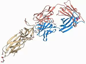
Background imageMolecular Structure Collection: Ebola virus glycoprotein and antibody F006 / 9738

Ebola virus glycoprotein and antibody F006 / 9738
Ebola virus glycoprotein and antibody. Molecular model of a surface glycoprotein from the Ebola virus (EBOV) bound to an antibody

Background imageMolecular Structure Collection: Urea channel molecule F006 / 9735

Urea channel molecule F006 / 9735
Urea channel. Molecular model of the proton-gated urea channel from the bacterium Helicobacter pylori. This channel opens in acidic environments, allowing the entry of urea into the cell

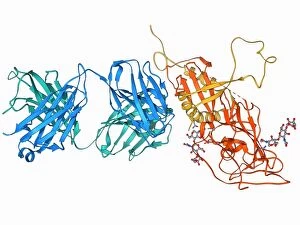
Background imageMolecular Structure Collection: Leptin receptor molecule F006 / 9736

Leptin receptor molecule F006 / 9736
Leptin receptor. Molecular model of a leptin receptor complexed with an antibody. Leptin is a hormone produced by adipose (fat) tissue

Background imageMolecular Structure Collection: DNA clamp complexed with DNA molecule F006 / 9732

DNA clamp complexed with DNA molecule F006 / 9732
DNA clamp complexed with DNA molecule. Molecular model showing a sliding DNA (deoxyribonucleic acid) clamp (ring) complexed with a molecule of DNA (blue and red)

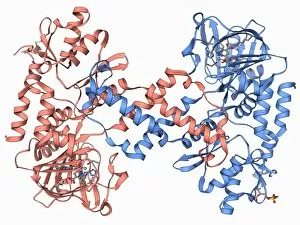
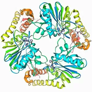
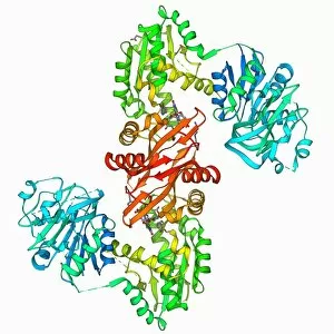
Background imageMolecular Structure Collection: GMP synthetase enzyme F006 / 9734

GMP synthetase enzyme F006 / 9734
GMP synthetase enzyme, molecular model. This enzyme, guanine monophosphate synthetase, catalyses the reaction that converts xanthosine monophosphate to guanosine monophosphate

Background imageMolecular Structure Collection: Single stranded DNA-binding protein F006 / 9733

Single stranded DNA-binding protein F006 / 9733
Single stranded DNA-binding protein (SSBP). Molecular model of a protein that binds to the single stranded DNA (deoxyribonucleic acid) in human mitochondria

Background imageMolecular Structure Collection: Sir3 gene silencer acting on DNA F006 / 9730

Sir3 gene silencer acting on DNA F006 / 9730
Sir3 gene silencer acting on DNA, molecular model. Sir3 (light blue) is acting on a circular strand of DNA (deoxyribonucleic acid, pink)

Background imageMolecular Structure Collection: Parathion hydrolase enzyme F006 / 9731

Parathion hydrolase enzyme F006 / 9731
Parathion hydrolase, molecular model. This enzyme hydrolyses bonds in organophosphates, which include pesticides and the nerve gas sarin

Background imageMolecular Structure Collection: Tumour suppressor protein with DNA F006 / 9729

Tumour suppressor protein with DNA F006 / 9729
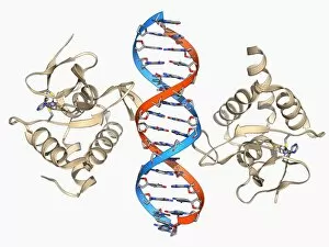
Tumour suppressor protein. Molecular model of the tumour suppressor protein p53 (beige) bound to a molecule of DNA (deoxyribonucleic acid, red and blue)

Background imageMolecular Structure Collection: Iron-regulatory protein bound to RNA F006 / 9727

Iron-regulatory protein bound to RNA F006 / 9727
Iron-regulatory protein bound to RNA, molecular model. Iron regulatory protein 1 (IRP1, purple) bound to a short strand of RNA (ribonucleic acid, red) that includes iron-responsive elements (IREs)

Background imageMolecular Structure Collection: DNA repair enzyme, molecular model F006 / 9726

DNA repair enzyme, molecular model F006 / 9726
DNA repair enzyme. Molecular model of the DNA (deoxyribonucleic acid) repair enzyme alpha-ketoglutarate-dependent dioxygenase bound to a molecule of DNA (red and blue)

Background imageMolecular Structure Collection: Oxidoreductase enzyme complex F006 / 9725

Oxidoreductase enzyme complex F006 / 9725
Oxidoreductase enzyme complex, molecular model. This is the membrane-bound domain formed from of a complex of NADH-quinone oxidoreductase subunits

Background imageMolecular Structure Collection: Saccharide transport protein F006 / 9722

Saccharide transport protein F006 / 9722
Saccharide transport protein. Molecular model of the phosphorylation-coupled saccharide transporter EIIC from the bacterium Bacillus cereus. EIIC is an integral membrane protein

Background imageMolecular Structure Collection: Aldo-keto reductase enzyme and ibuprofen F006 / 9724

Aldo-keto reductase enzyme and ibuprofen F006 / 9724
Aldo-keto reductase enzyme and ibuprofen. Molecular model of the enzyme aldo-keto reductase family 1 member C3 (AKR1C3) bound to a molecule of the anti-inflammatory drug ibuprofen

Background imageMolecular Structure Collection: SMAD4 protein domain bound to DNA F006 / 9723

SMAD4 protein domain bound to DNA F006 / 9723
SMAD4 protein domain bound to DNA, molecular model. This strand of DNA (deoxyribonucleic acid, red and blue) is surrounded by MH1 domains of the SMAD4 (Mothers against decapentaplegic homolog 4)

Background imageMolecular Structure Collection: Transcription activator and DNA F006 / 9721

Transcription activator and DNA F006 / 9721
Transcription activator and DNA. Molecular model of the transcriptional activator of the multidrug efflux transporter BmrR bound to a molecule of DNA (deoxyribonucleic acid, red and blue)

Background imageMolecular Structure Collection: Methyltransferase complexed with DNA F006 / 9711

Methyltransferase complexed with DNA F006 / 9711
Methyltransferase complexed with DNA, molecular model. The strand of DNA (deoxyribonucleic acid, red and blue) is enclosed by DNA methyltransferase 1 (DNMT-1, beige)

Background imageMolecular Structure Collection: Pepsinogen molecule F006 / 9710

Pepsinogen molecule F006 / 9710
Pepsinogen. Molecular model of pepsinogen, the inactive precursor to the digestive enzyme pepsin. Pepsion, which is released by the stomach digests proteins

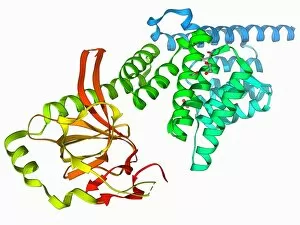
Background imageMolecular Structure Collection: Glycosylation enzyme molecule F006 / 9708

Glycosylation enzyme molecule F006 / 9708
Glycosylation enzyme. Molecular model of the enzyme N-acetylglucosamine (GlcNAc) transferase. This intracellular enzyme adds N-acetylglucosamine molecules to target proteins


All Professionally Made to Order for Quick Shipping

"Molecular Structure: Unlocking the Secrets of Life's Building Blocks" From anaesthetics inhibiting ion channels to antidepressant molecules, the intricate world holds endless wonders. The C015 / 6718 anaesthetic molecule delicately interacts with ion channels, altering their function and providing relief from pain. Meanwhile, Amitriptyline, an antidepressant molecule, works its magic by modulating neurotransmitters in our brains. In the realm of immunity, Immunoglobulin G antibody F007 / 9894 stands tall as a defender against pathogens. Its unique structure allows it to recognize and neutralize foreign invaders effectively. On another front, DNA artwork showcases the elegance and complexity that underlies all life forms on Earth. Creatine amino acid molecule fuels our muscles during intense physical activities while nanotube technology revolutionizes various industries with its exceptional properties. These tiny tubes hold immense potential for advancements in medicine and materials science alike. Zinc fingers bound to a DNA strand demonstrate how proteins can precisely interact with genetic material. This interaction plays a crucial role in gene regulation and expression. Carbon nanotubes take center stage once again as they exhibit remarkable strength and conductivity at the nano-scale level. Oxytocin neurotransmitter molecule reminds us of love's powerful influence on human connections—its presence promotes bonding between individuals. Manganese superoxide dismutase enzyme F006 / 9423 safeguards our cells by combating harmful free radicals that contribute to aging and disease. Even viruses have their own molecular structures; SARS coronavirus protein represents one such example—a key player in viral replication within host cells. Conceptual artwork further explores nanotube technology's limitless possibilities—the fusion of imagination and scientific innovation knows no bounds here. As we delve deeper into understanding molecular structures, we unravel nature's blueprint for life itself—one atom at a time. These captivating glimpses into the microscopic world remind us of both the fragility and resilience found within the building blocks of existence.