Fraud Blocker Skip to main content

Molecular Structure Collection (page 15)

"Molecular Structure: Unlocking the Secrets of Life's Building Blocks" From anaesthetics inhibiting ion channels to antidepressant molecules

Background imageMolecular Structure Collection: Androgen receptor and modulator F006 / 9660

Androgen receptor and modulator F006 / 9660
Androgen receptor and modulator. Molecular model of a selective androgen receptor modulator (SARM) bound to an androgen receptor

Background imageMolecular Structure Collection: Bacterial biofilm enzyme F006 / 9658

Bacterial biofilm enzyme F006 / 9658
Bacterial biofilm enzyme. Molecular model of the enzyme glucansucrase from the bacterium Streptococcus mutans. This enzyme is used to form bacterial biofilms on teeth that cause dental caries

Background imageMolecular Structure Collection: Rous sarcoma virus capsid F006 / 9653

Rous sarcoma virus capsid F006 / 9653
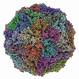
Rous sarcoma virus capsid, molecular model. In viruses, the capsid is the protein shell that encloses the genetic material

Background imageMolecular Structure Collection: Rabbit eye lens protein molecule F006 / 9657

Rabbit eye lens protein molecule F006 / 9657
Rabbit eye lens protein. Molecular model of lambda-crystallin, a structural protein found in the eye lenses of rabbits (family Leporidae)

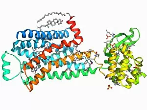
Background imageMolecular Structure Collection: Sodium-potassium ion pump protein F006 / 9656

Sodium-potassium ion pump protein F006 / 9656
Sodium-potassium ion pump protein, molecular model. Sodium-potassium ATPase (adenosine triphosphatase) is an ATP-powered ion pump found in all animal cells

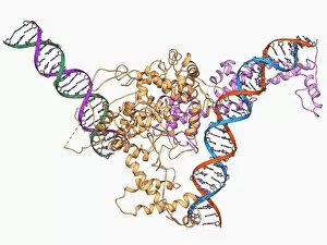
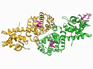
Background imageMolecular Structure Collection: Chromatin remodelling factor and DNA F006 / 9655

Chromatin remodelling factor and DNA F006 / 9655
Chromatin remodelling factor and DNA, molecular model. The strands of DNA (deoxyribonucleic acid) are at left and right. This chromatin remodelling factor is ISW1a

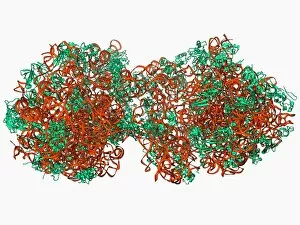
Background imageMolecular Structure Collection: 70S ribosome, molecular model F006 / 9651

70S ribosome, molecular model F006 / 9651
70S ribosome, molecular model. Ribosomes are composed of protein and RNA (ribonucleic acid). In bacteria each ribosome consists of a small (30S) subunit and a large (50S) subunit

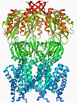
Background imageMolecular Structure Collection: MscS ion channel protein structure F006 / 9650

MscS ion channel protein structure F006 / 9650
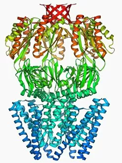
MscS ion channel protein structure. Molecular model of a mechanosensitive channel of small conductance (MscS) from an Escherichia coli bacterium

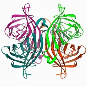
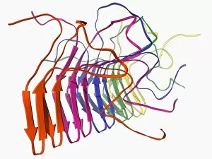
Background imageMolecular Structure Collection: Streptavidin bacterial protein F006 / 9654

Streptavidin bacterial protein F006 / 9654
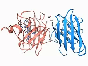
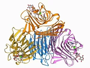
Streptavidin bacterial protein, molecular model. Streptavidin is a protein obtained from the bacterium Streptomyces avidinii

Background imageMolecular Structure Collection: Fatty acid synthase molecule F006 / 9647

Fatty acid synthase molecule F006 / 9647
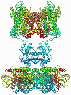
Fatty acid synthase (FAS), molecular model. FAS is a multi-enzyme that plays a key role in the synthesis of fatty acids (lipids) in the human body. It is not a single enzyme but a whole enzyme system

Background imageMolecular Structure Collection: Eye lens protein molecule F006 / 9652

Eye lens protein molecule F006 / 9652
Eye lens protein. Molecular model of alpha-crystallin, a protein found in the lens of the eye. The regular arrangement of the protein in the lens is thought to be responsible for its transparency

Background imageMolecular Structure Collection: Integrin and fibrinogen complex molecule F006 / 9649

Integrin and fibrinogen complex molecule F006 / 9649
Integrin and fibrinogen complex. Molecular model of integrin alpha-II beta-3 complexed with fibrinogen. Integrin alpha-II beta-3 is a transmembrane protein found on platelets

Background imageMolecular Structure Collection: Grapevine fanleaf virus capsid

Grapevine fanleaf virus capsid, molecular model. This plant virus is named for its infection of grape vines. It is transmitted by the nematode worm Xiphinema index

Background imageMolecular Structure Collection: SelB elongation factor bound to RNA F006 / 9648

SelB elongation factor bound to RNA F006 / 9648
SelB elongation factor bound to RNA. Molecular model of the SelB elongation factor bound to an mRNA (messenger ribonucleic acid) hairpin formed by the selenocysteine insertion sequence (SECIS)

Background imageMolecular Structure Collection: Voltage-gated potassium channel F006 / 9642

Voltage-gated potassium channel F006 / 9642
Voltage-gated potassium channel. Molecular model of a voltage-gated potassium (Kv) ion channel. Ion channels are membrane-spanning proteins that form pores in cell membranes

Background imageMolecular Structure Collection: Src protein molecule F006 / 9646

Src protein molecule F006 / 9646
Src protein, molecular model. Src is a tyrosine kinase, a signalling protein in cells that has the ability to turn on protein synthesis and cellular growth

Background imageMolecular Structure Collection: Copper, zinc superoxide dismutase enzyme F006 / 9645

Copper, zinc superoxide dismutase enzyme F006 / 9645
Copper, zinc superoxide dismutase enzyme

Background imageMolecular Structure Collection: Fungal prion protein F006 / 9644

Fungal prion protein F006 / 9644
Fungal prion protein. Molecular model of the amyloid form of the HET prion protein

Background imageMolecular Structure Collection: Beta-2 adrenergic receptor molecule F006 / 9643

Beta-2 adrenergic receptor molecule F006 / 9643
Beta-2 adrenergic receptor. Molecular model of a human beta-2 adrenergic receptor bound to an antibody. Beta receptors respond to adrenalin, causing a sympathetic (fight or flight) response

Background imageMolecular Structure Collection: Adenovirus protein and tumor suppressor F006 / 9641

Adenovirus protein and tumor suppressor F006 / 9641
Adenovirus protein and tumour suppressor. Molecular model of the E1A protein from human adenovirus bound to a retinoblastoma tumour suppressor

Background imageMolecular Structure Collection: Beta-2 adrenergic receptor molecule F006 / 9640

Beta-2 adrenergic receptor molecule F006 / 9640
Beta-2 adrenergic receptor. Molecular model of a human beta-2 adrenergic receptor bound to an antibody. Beta receptors respond to adrenalin, causing a sympathetic (fight or flight) response

Background imageMolecular Structure Collection: E coli virulence factor F006 / 9639

E coli virulence factor F006 / 9639
E. coli virulence factor. Molecular model of the beta-domain of the EspP autotransporter protein from the bacterium Escherichia coli

Background imageMolecular Structure Collection: 70S ribosome, molecular model F006 / 9638

70S ribosome, molecular model F006 / 9638
70S ribosome. Molecular model of a 70S ribosome complex containing a Shine-Dalgarno helix, the point of mRNA (messenger ribonucleic acid) binding

Background imageMolecular Structure Collection: Metal-sensing RNA molecule F006 / 9636

Metal-sensing RNA molecule F006 / 9636
Metal-sensing RNA molecule. Molecular model of an M-box riboswitch, a length of RNA (ribonucleic acid) that regulates levels of metal ions in a cell

Background imageMolecular Structure Collection: Trypsin molecule F006 / 9634

Trypsin molecule F006 / 9634
Trypsin molecule. Molecular model of the digestive protease enzyme trypsin. Trypsin is released by the pancreas to break down proteins into smaller chains of amino acids

Background imageMolecular Structure Collection: PolyA polymerase and RNA F006 / 9635

PolyA polymerase and RNA F006 / 9635
Poly(A) polymerase and RNA. Molecular model of poly(A) polymerase complexed with RNA (ribonucleic acid) and ATP (adenosine triphosphate)

Background imageMolecular Structure Collection: Internal ribosome entry site F006 / 9631

Internal ribosome entry site F006 / 9631
Internal ribosome entry site. Molecular model of an internal ribosome entry site nucleotide sequence from the hepatitis C virus. This sequence is essential for the initiation of viral translation

Background imageMolecular Structure Collection: Trypsin molecule with inhibitor F006 / 9633

Trypsin molecule with inhibitor F006 / 9633
Trypsin molecule. Molecular model of the digestive protease enzyme beta-trypsin complexed with an inhibitor. Trypsin is released by the pancreas to break down proteins into smaller chains of amino

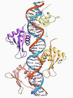
Background imageMolecular Structure Collection: Interferon regulatory factor molecule F006 / 9630

Interferon regulatory factor molecule F006 / 9630
Interferon regulatory factor. Molecular model of interferon regulatory factor 3 (IRF3, coils at right and left) bound to a DNA (deoxyribonucleic acid) molecule (red and blue)

Background imageMolecular Structure Collection: Wilms tumor suppressor bound to DNA F006 / 9632

Wilms tumor suppressor bound to DNA F006 / 9632
Wilms tumour suppressor bound to DNA. Molecular model of the zinc finger domain of the Wilms tumour suppressor protein bound to a strand of DNA (deoxyribonucleic acid)

Background imageMolecular Structure Collection: Lactose binding protein molecule F006 / 9629

Lactose binding protein molecule F006 / 9629
Lactose binding protein. Molecular model of a lectin protein from the peanut plant (Arachis hypogaea) bound to a lactose molecule

Background imageMolecular Structure Collection: MscS ion channel protein structure F006 / 9626

MscS ion channel protein structure F006 / 9626
MscS ion channel protein structure. Molecular model of a mechanosensitive channel of small conductance (MscS) from an Escherichia coli bacterium

Background imageMolecular Structure Collection: Transport inhibitor response 1 protein F006 / 9628

Transport inhibitor response 1 protein F006 / 9628
Transport inhibitor response 1 protein. Molecular model of the transport inhibitor response 1 protein bound to the plant hormone auxin. This protein is involved in auxin gene regulation

Background imageMolecular Structure Collection: Multidrug transporter molecule F006 / 9627

Multidrug transporter molecule F006 / 9627
Multidrug transporter. Molecular model of the multidrug transporter Sav1866 from the bacterium Escherichia coli. This protein pumps drugs, including antibiotics, out of the bacterial cell

Background imageMolecular Structure Collection: Mengovirus capsid, molecular model F006 / 9617

Mengovirus capsid, molecular model F006 / 9617
Mengovirus capsid, molecular model. A capsid consists of subunits called capsomeres that self-assemble to form the shell seen here

Background imageMolecular Structure Collection: Insulin molecule F006 / 9625

Insulin molecule F006 / 9625
Insulin, molecular model. Insulin plays an important role in blood sugar regulation. It is released from the pancreas when blood sugar levels are high, for example after a meal

Background imageMolecular Structure Collection: MscL ion channel protein structure F006 / 9624

MscL ion channel protein structure F006 / 9624
MscL ion channel protein structure. Molecular model of a mechanosensitive channel of large conductance (MscL) from a Mycobacterium tuberculosis bacterium

Background imageMolecular Structure Collection: Endoplasmic reticulum chaperone protein F006 / 9623

Endoplasmic reticulum chaperone protein F006 / 9623
Endoplasmic reticulum chaperone protein. Molecular model of the endoplasmic reticulum chaperone protein GRP94. This protein is essential for the maturation of cell-surface display proteins

Background imageMolecular Structure Collection: RNA exosome complex, molecular model F006 / 9620

RNA exosome complex, molecular model F006 / 9620
RNA exosome complex, molecular model. This multi-protein complex functions to break up strands of RNA (ribonucleic acid, pink) during biochemical processes

Background imageMolecular Structure Collection: HIV antibody therapy, molecular model F006 / 9622

HIV antibody therapy, molecular model F006 / 9622
HIV antibody therapy. Molecular model of the interaction of the HIV surface protein gp120 (green) as it interacts with a human white blood cell surface protein (CD4)

Background imageMolecular Structure Collection: Rhomboid protease molecule F006 / 9621

Rhomboid protease molecule F006 / 9621
Rhomboid protease. Molecular model of the rhomboid protease enzyme GlpG from the bacterium Escherichia coli. Proteases are enzymes that break down proteins

Background imageMolecular Structure Collection: Kinesin motor protein F006 / 9619

Kinesin motor protein F006 / 9619
Kinesin motor protein. Molecular model of the ncd kinesin motor protein. Kinesin motor proteins transport vesicles containing intracellular cargo around the cell along microtubules

Background imageMolecular Structure Collection: Molecular motor protein F006 / 9618

Molecular motor protein F006 / 9618
Myosin molecular motor protein, molecular model. Motor proteins convert chemical energy into mechanical movements in response to specific chemical stimuli

Background imageMolecular Structure Collection: RNA editing enzyme F006 / 9615

RNA editing enzyme F006 / 9615
RNA editing enzyme, molecular model. This enzyme binds to double-stranded RNA (ribonucleic acid)

Background imageMolecular Structure Collection: Lysozyme molecule F006 / 9616

Lysozyme molecule F006 / 9616
Lysozyme, molecular model. Lysozymes are enzymes found in a wide range of biological fluids such as tears, saliva and milk. This lysozyme is from chicken egg white

Background imageMolecular Structure Collection: RNA-dependent RNA polymerase molecule F006 / 9611

RNA-dependent RNA polymerase molecule F006 / 9611
RNA-dependent RNA polymerase, molecular model. This enzyme catalyses the replication of RNA (ribonucleic acid) from an RNA template

Background imageMolecular Structure Collection: Titin muscle protein molecule F006 / 9612

Titin muscle protein molecule F006 / 9612
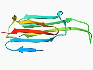
Titin muscle protein. Molecular model of two immunoglobulin-like domains from the giant muscle protein titin

Background imageMolecular Structure Collection: Integrin transmembrane domain F006 / 9614

Integrin transmembrane domain F006 / 9614
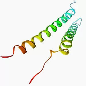
Integrin transmembrane domain, molecular model. Integrins are transmembrane cell adhesion receptors


All Professionally Made to Order for Quick Shipping

"Molecular Structure: Unlocking the Secrets of Life's Building Blocks" From anaesthetics inhibiting ion channels to antidepressant molecules, the intricate world holds endless wonders. The C015 / 6718 anaesthetic molecule delicately interacts with ion channels, altering their function and providing relief from pain. Meanwhile, Amitriptyline, an antidepressant molecule, works its magic by modulating neurotransmitters in our brains. In the realm of immunity, Immunoglobulin G antibody F007 / 9894 stands tall as a defender against pathogens. Its unique structure allows it to recognize and neutralize foreign invaders effectively. On another front, DNA artwork showcases the elegance and complexity that underlies all life forms on Earth. Creatine amino acid molecule fuels our muscles during intense physical activities while nanotube technology revolutionizes various industries with its exceptional properties. These tiny tubes hold immense potential for advancements in medicine and materials science alike. Zinc fingers bound to a DNA strand demonstrate how proteins can precisely interact with genetic material. This interaction plays a crucial role in gene regulation and expression. Carbon nanotubes take center stage once again as they exhibit remarkable strength and conductivity at the nano-scale level. Oxytocin neurotransmitter molecule reminds us of love's powerful influence on human connections—its presence promotes bonding between individuals. Manganese superoxide dismutase enzyme F006 / 9423 safeguards our cells by combating harmful free radicals that contribute to aging and disease. Even viruses have their own molecular structures; SARS coronavirus protein represents one such example—a key player in viral replication within host cells. Conceptual artwork further explores nanotube technology's limitless possibilities—the fusion of imagination and scientific innovation knows no bounds here. As we delve deeper into understanding molecular structures, we unravel nature's blueprint for life itself—one atom at a time. These captivating glimpses into the microscopic world remind us of both the fragility and resilience found within the building blocks of existence.