Fraud Blocker Skip to main content

Molecular Structure Collection (page 17)

"Molecular Structure: Unlocking the Secrets of Life's Building Blocks" From anaesthetics inhibiting ion channels to antidepressant molecules

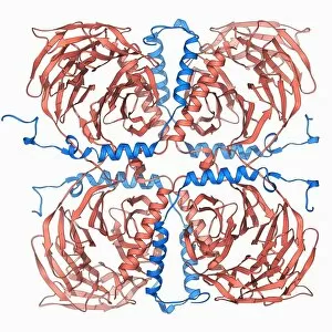
Background imageMolecular Structure Collection: Ricin molecule F006 / 9563

Ricin molecule F006 / 9563
Ricin. Molecular model of the toxic protein ricin. It comprises two entwined amino acid chains, termed A and B. The A-chain is toxic, inhibiting protein synthesis in cells

Background imageMolecular Structure Collection: DNA tetranucleosome, molecular model F006 / 9555

DNA tetranucleosome, molecular model F006 / 9555
DNA tetranucleosome. Molecular model of four nucleosomes, or a tetranucleosome. Nucleosomes are the fundamental repeating unit used to package DNA (deoxyribonucleic acid) inside cell nuclei
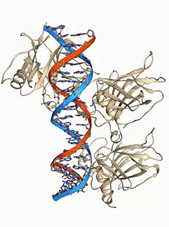

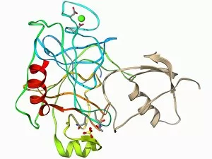
Background imageMolecular Structure Collection: Thrombin complexed with fibrinogen F006 / 9561

Thrombin complexed with fibrinogen F006 / 9561
Thrombin complexed with fibrinogen, molecular model. The thrombin molecules (beige, right and left) are bound to the central part of the fibrinogen molecule (down centre)

Background imageMolecular Structure Collection: SARS virus surface protein molecule F006 / 9560

SARS virus surface protein molecule F006 / 9560
SARS virus surface protein. Molecular model of the spike 2 (S2) envelope glycoprotein from the severe acute respiratory syndrome (SARS) virus

Background imageMolecular Structure Collection: DNA polymerase with DNA F006 / 9559

DNA polymerase with DNA F006 / 9559
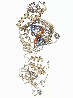
DNA polymerase with DNA. Molecular model of human DNA polymerase beta (beige) complexed with a molecule of DNA (deoxyribonucleic acid, red and blue)

Background imageMolecular Structure Collection: Insulin molecule F006 / 9558

Insulin molecule F006 / 9558
Insulin, molecular model. Insulin plays an important role in blood sugar regulation. It is released from the pancreas when blood sugar levels are high, for example after a meal

Background imageMolecular Structure Collection: Bacteriophage DNA recombination F006 / 9554

Bacteriophage DNA recombination F006 / 9554
Bacteriophage DNA recombination. Molecular model showing DNA manipulation and recombination taking place at a Holliday junction with a bacteriophage enzyme

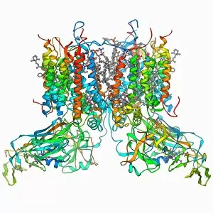
Background imageMolecular Structure Collection: ATP-dependent protease molecule F006 / 9552

ATP-dependent protease molecule F006 / 9552
ATP-dependent protease. Molecular model of the bacterial enzyme HsIUV protease. Proteases are enzymes that break down proteins. HsIUV is expressed in response to cellular stress

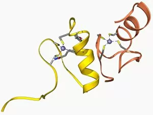
Background imageMolecular Structure Collection: Zinc finger molecule F006 / 9557

Zinc finger molecule F006 / 9557
Zinc finger, molecular model. Zinc fingers constitute the DNA recognition domains of many DNA regulatory proteins and are so named for their resemblance to fingers projecting from the protein

Background imageMolecular Structure Collection: Botulinum type B neurotoxin F006 / 9553

Botulinum type B neurotoxin F006 / 9553
Botulinum type B neurotoxin, molecular model. This powerful toxin, produced by the bacterium Clostridium botulinum, enters nerve cells and prevents release of neurotransmitters

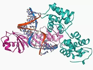
Background imageMolecular Structure Collection: TATA box-binding protein complex F006 / 9551

TATA box-binding protein complex F006 / 9551
TATA box-binding protein complex. Molecular model showing a yeast TATA box-binding protein (TBP) complexed with a strand of DNA (deoxyribonucleic acid, red and blue) and transcription factor IIA

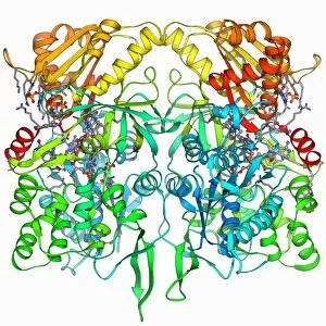
Background imageMolecular Structure Collection: NADP-dependent alcohol dehydrogenase F006 / 9549

NADP-dependent alcohol dehydrogenase F006 / 9549
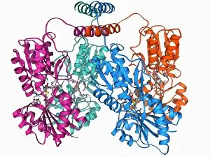
NADP-dependent alcohol dehydrogenase, molecular model. Alcohol dehydrogenase (ADH) is an enzyme that facilitates the break-down of alcohols in the body, which could otherwise be toxic

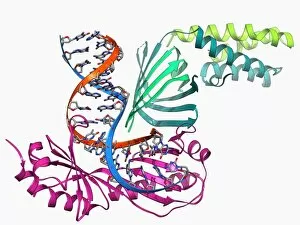
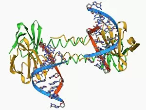
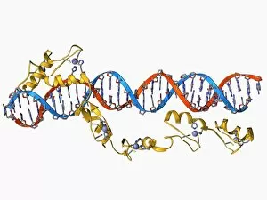
Background imageMolecular Structure Collection: TATA box-binding protein and DNA F006 / 9550

TATA box-binding protein and DNA F006 / 9550
TATA box-binding protein and DNA. Molecular model showing a TATA box-binding protein (TBP) complexed with a strand of DNA (deoxyribonucleic acid, red and blue)

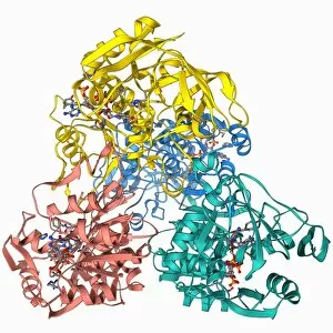
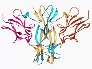
Background imageMolecular Structure Collection: Cholera toxin molecule F006 / 9546

Cholera toxin molecule F006 / 9546
Cholera toxin, molecular model. Cholera toxin is a protein complex secreted by the bacterium Vibrio cholerae, that causes the disease cholera

Background imageMolecular Structure Collection: Programmed cell death protein 6 molecule F006 / 9548

Programmed cell death protein 6 molecule F006 / 9548
Programmed cell death protein 6, molecular model. This is a calcium-binding protein is involved in apoptosis (programmed cell death)

Background imageMolecular Structure Collection: GATA transcription factor and zinc finger F006 / 9547

GATA transcription factor and zinc finger F006 / 9547
GATA transcription factor. Molecular model of the GATA transcription factor bound to a zinc finger. Transcription factors are proteins that bind to specific DNA sequences

Background imageMolecular Structure Collection: Plant hormone regulator, molecular model F006 / 9545

Plant hormone regulator, molecular model F006 / 9545
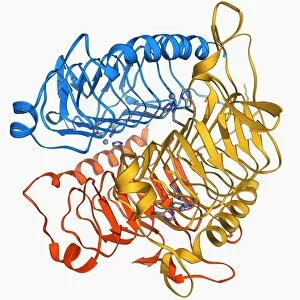
Plant hormone regulator. Molecular model of Ia-amidohydrolase from thale cress (Arabidopsis thaliana). This enzyme acts to release the plant hormone indoe-3-acetic acid from its storage form

Background imageMolecular Structure Collection: Hydroxysteroid dehydrogenase molecule F006 / 9543

Hydroxysteroid dehydrogenase molecule F006 / 9543
Hydroxysteroid dehydrogenase. Molecular model of the human type 5 hydroxysteroid dehydrogenase enzyme bound to a molecule of the steroid drug androstene

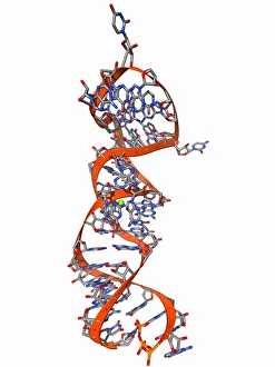
Background imageMolecular Structure Collection: RNA stem-loop motif, molecular model F006 / 9544

RNA stem-loop motif, molecular model F006 / 9544
RNA stem-loop motif. Molecular model of the stem-loop II motif from the SARS (severe acute respiratory syndrome) coronavirus. This RNA (ribonucleic acid) element is a target for antiviral drugs

Background imageMolecular Structure Collection: DNA 6-way junction, artwork C014 / 2585

DNA 6-way junction, artwork C014 / 2585
DNA (deoxyribonucleic acid) cube, computer artwork. The DNA cube is formed from six different DNA strands. Each side of the cube is composed of a single circular DNA strand

Background imageMolecular Structure Collection: Nerve growth factor bound to receptor F006 / 9541

Nerve growth factor bound to receptor F006 / 9541
Nerve growth factor bound to receptor, molecular model. Human nerve growth factor bound to the TrkA receptor. NGF is a neurotrophin that acts on the development and function of nerves

Background imageMolecular Structure Collection: DNA binding protein, molecular model F006 / 9540

DNA binding protein, molecular model F006 / 9540
DNA binding protein. Molecular model of the recombinant protein S7dLZ bound to DNA (deoxyribonucleic acid) molecules. S7dLZ is made up of the DNA binding protein Sac7d

Background imageMolecular Structure Collection: Pyruvate dehydrogenase enzyme molecule F006 / 9538

Pyruvate dehydrogenase enzyme molecule F006 / 9538
Pyruvate dehydrogenase (E1), molecular model. This enzyme converts pyruvate to acetyl-CoA for use in the citric acid (or Krebs) cycle

Background imageMolecular Structure Collection: Elongation factor Tu and tRNA F006 / 9522

Elongation factor Tu and tRNA F006 / 9522
Elongation factor Tu bound to tRNA (transfer ribonucleic acid), molecular model. This enzyme is involved in the elongation of polypeptide chains during translation

Background imageMolecular Structure Collection: Fish antifreeze protein F006 / 9539

Fish antifreeze protein F006 / 9539
Fish antifreeze protein. Molecular model of a fish antifreeze protein (AFP) from the winter flounder (Pseudopleuronectes americanus)

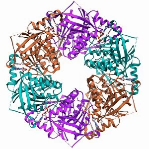
Background imageMolecular Structure Collection: Bacteriophage ATPase molecule F006 / 9536

Bacteriophage ATPase molecule F006 / 9536
Bacteriophage ATPase. Molecular model of an ATP synthase (ATPase) molecule from the phi 12 bacteriophage. ATPase is an important enzyme that provides energy for cells through the synthesis of

Background imageMolecular Structure Collection: Molecular motor protein F006 / 9537

Molecular motor protein F006 / 9537
Molecular motor protein. Molecular model of a two-headed motor protein, Myosin V. Motor proteins convert chemical energy into mechanical movements in response to specific chemical stimuli

Background imageMolecular Structure Collection: Cytochrome b6f complex, molecular model F006 / 9533

Cytochrome b6f complex, molecular model F006 / 9533
Cytochrome b6f complex, molecular model. Cytochrome molecules perform oxidation and reduction reactions for electron transport

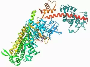
Background imageMolecular Structure Collection: Glycosylation enzyme molecule F006 / 9535

Glycosylation enzyme molecule F006 / 9535
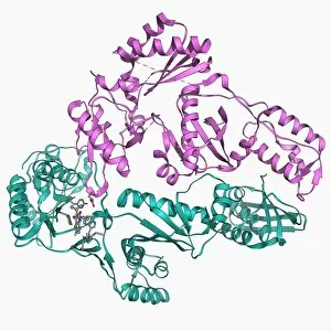
Glycosylation enzyme. Molecular model of the enzyme N-acetylglucosamine (GlcNAc) transferase. This intracellular enzyme adds N-acetylglucosamine molecules to target proteins

Background imageMolecular Structure Collection: TATA box-binding protein complex F006 / 9534

TATA box-binding protein complex F006 / 9534
TATA box-binding protein complex. Molecular model showing a yeast TATA box-binding protein (TBP) complexed with a strand of DNA (deoxyribonucleic acid, red and blue) and transcription factor IIB

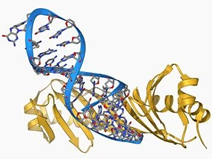
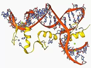
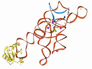
Background imageMolecular Structure Collection: Transcription factor and ribosomal RNA F006 / 9530

Transcription factor and ribosomal RNA F006 / 9530
Transcription factor and ribosomal RNA (rRNA). Molecular model showing the 6 zinc fingers of transcription factor IIIA (yellow) bound to RNA (ribonucleic acid)

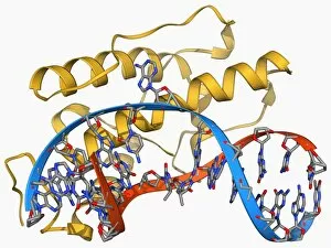
Background imageMolecular Structure Collection: Bacteriophage restriction enzyme F006 / 9531

Bacteriophage restriction enzyme F006 / 9531
Bacteriophage restriction enzyme. Molecular model of the restriction enzyme endonuclease V (yellow) from the bacteriophage T4 complexed with DNA (deoxyribonucleic acid, red and blue)

Background imageMolecular Structure Collection: Squalene-hopene cyclase molecule F006 / 9529

Squalene-hopene cyclase molecule F006 / 9529
Squalene-hopene cyclase, molecular model. This bacterial enzyme catalyses the cyclization of squalene to hopene

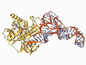
Background imageMolecular Structure Collection: Self-splicing RNA intron, molecular model F006 / 9527

Self-splicing RNA intron, molecular model F006 / 9527
Self-splicing RNA intron, molecular model. Splicing is the process where a non-coding fragment (intron) of a strand of nucleic acid (DNA, deoxyribonucleic acid; or RNA, ribonucleic acid) is removed

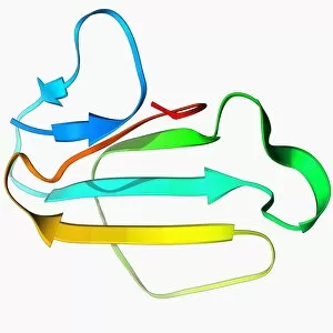
Background imageMolecular Structure Collection: Ubiquitin molecule F006 / 9528

Ubiquitin molecule F006 / 9528
Ubiquitin, molecular model. Ubiquitin is found in all eukaryotic cells. When a protein is damaged or old it will be tagged by several ubiquitin molecules

Background imageMolecular Structure Collection: Cobra venom molecule F006 / 9524

Cobra venom molecule F006 / 9524
Cobra venom. Molecular model of toxin b, a long neurotoxin from a king cobra (Ophiophagus hannah)

Background imageMolecular Structure Collection: Tumour suppressor protein with DNA F006 / 9523

Tumour suppressor protein with DNA F006 / 9523
Tumour suppressor protein. Molecular model of the tumour suppressor protein p53 (beige) bound to a molecule of DNA (deoxyribonucleic acid, red and blue)

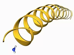
Background imageMolecular Structure Collection: Tarantula toxin molecule F006 / 9525

Tarantula toxin molecule F006 / 9525
Tarantula toxin. Molecular model of a peptide toxin from the tarantula Grammostola spatulata. This toxin works by inhibiting mechanosensitive ion channels

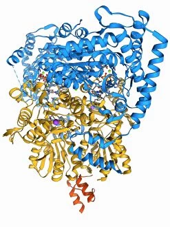
Background imageMolecular Structure Collection: Nitric oxide synthase molecule F006 / 9521

Nitric oxide synthase molecule F006 / 9521
Nitric oxide synthase, molecular model. This enzyme catalyses the production of nitric oxide from L-arginine. Nitric oxide is involved in cellular signalling

Background imageMolecular Structure Collection: LAC repressor molecule F006 / 9520

LAC repressor molecule F006 / 9520
LAC repressor. Molecular model of a LAC (lactose) repressor molecule. The LAC repressor inhibits the expression of genes that code for an enzyme which metabolizes lactose in bacteria

Background imageMolecular Structure Collection: SV40 virus capsid, molecular model F006 / 9508

SV40 virus capsid, molecular model F006 / 9508
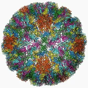
SV40 virus capsid, molecular model. Simian virus 40 (SV40) is found in monkeys such as Rhesus monkeys and macaques. Potentially tumour-causing, it is used in laboratory research and in vaccines

Background imageMolecular Structure Collection: Carbonic anhydrase molecule F006 / 9518

Carbonic anhydrase molecule F006 / 9518
Carbonic anhydrase, molecular model. This enzyme catalyses the reversible hydration of carbon dioxide

Background imageMolecular Structure Collection: Reverse transcriptase and inhibitor F006 / 9519

Reverse transcriptase and inhibitor F006 / 9519
Reverse transcriptase and inhibitor. Molecular model of HIV reverse transcriptase complexed with a non-nucleoside reverse transcriptase inhibitor drug

Background imageMolecular Structure Collection: Transducin protein beta-gamma complex F006 / 9514

Transducin protein beta-gamma complex F006 / 9514
Transducin protein beta-gamma complex. Molecular model of the beta-gamma dimer of the heterotrimeric G protein transducin

Background imageMolecular Structure Collection: Trypsinogen molecule with inhibitor F006 / 9517

Trypsinogen molecule with inhibitor F006 / 9517
Trypsinogen molecule. Molecular model of trypsinogen, the precursor to the digestive protease enzyme trypsin, complexed with an inhibitor

Background imageMolecular Structure Collection: Transcription factor and ribosomal RNA F006 / 9516

Transcription factor and ribosomal RNA F006 / 9516
Transcription factor and ribosomal RNA (rRNA). Molecular model showing the 6 zinc fingers of transcription factor IIIA (yellow) bound to RNA (ribonucleic acid)

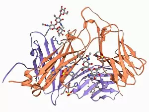
Background imageMolecular Structure Collection: T cell receptor, molecular model F006 / 9515

T cell receptor, molecular model F006 / 9515
T cell receptor. Molecular model of an alpha T cell receptor. T cell receptors are protein complexes found on the surface of a type of white blood cell called T lymphocytes (or T cells)

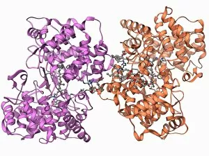
Background imageMolecular Structure Collection: DNA polymerase with DNA F006 / 9512

DNA polymerase with DNA F006 / 9512
DNA polymerase with DNA. Molecular model of DNA polymerase (purple) complexed with a molecule of DNA (deoxyribonucleic acid, pink and blue)


All Professionally Made to Order for Quick Shipping

"Molecular Structure: Unlocking the Secrets of Life's Building Blocks" From anaesthetics inhibiting ion channels to antidepressant molecules, the intricate world holds endless wonders. The C015 / 6718 anaesthetic molecule delicately interacts with ion channels, altering their function and providing relief from pain. Meanwhile, Amitriptyline, an antidepressant molecule, works its magic by modulating neurotransmitters in our brains. In the realm of immunity, Immunoglobulin G antibody F007 / 9894 stands tall as a defender against pathogens. Its unique structure allows it to recognize and neutralize foreign invaders effectively. On another front, DNA artwork showcases the elegance and complexity that underlies all life forms on Earth. Creatine amino acid molecule fuels our muscles during intense physical activities while nanotube technology revolutionizes various industries with its exceptional properties. These tiny tubes hold immense potential for advancements in medicine and materials science alike. Zinc fingers bound to a DNA strand demonstrate how proteins can precisely interact with genetic material. This interaction plays a crucial role in gene regulation and expression. Carbon nanotubes take center stage once again as they exhibit remarkable strength and conductivity at the nano-scale level. Oxytocin neurotransmitter molecule reminds us of love's powerful influence on human connections—its presence promotes bonding between individuals. Manganese superoxide dismutase enzyme F006 / 9423 safeguards our cells by combating harmful free radicals that contribute to aging and disease. Even viruses have their own molecular structures; SARS coronavirus protein represents one such example—a key player in viral replication within host cells. Conceptual artwork further explores nanotube technology's limitless possibilities—the fusion of imagination and scientific innovation knows no bounds here. As we delve deeper into understanding molecular structures, we unravel nature's blueprint for life itself—one atom at a time. These captivating glimpses into the microscopic world remind us of both the fragility and resilience found within the building blocks of existence.