Fraud Blocker Skip to main content

Molecular Structure Collection (page 18)

"Molecular Structure: Unlocking the Secrets of Life's Building Blocks" From anaesthetics inhibiting ion channels to antidepressant molecules

Background imageMolecular Structure Collection: DNA polymerase with DNA F006 / 9512

DNA polymerase with DNA F006 / 9512
DNA polymerase with DNA. Molecular model of DNA polymerase (purple) complexed with a molecule of DNA (deoxyribonucleic acid, pink and blue)

Background imageMolecular Structure Collection: DNA helicase molecule F006 / 9509

DNA helicase molecule F006 / 9509
DNA helicase. Molecular model of a helicase molecule from the SV40 virus. Helicases are enzymes that separate the two strands of the DNA double helix

Background imageMolecular Structure Collection: Simian virus SV40 large T antigen F006 / 9513

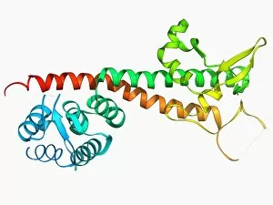
Simian virus SV40 large T antigen F006 / 9513
Simian virus (SV40) large T antigen, molecular model. This antigen is from the simian vacuolating virus 40 (SV40). Large T antigens play a role in regulating the viral life cycle of

Background imageMolecular Structure Collection: Transcription activation of IFN-beta gene F006 / 9510

Transcription activation of IFN-beta gene F006 / 9510
Transcription activation of IFN-beta gene. Molecular model of an enhanceosome containing the transcription factors IRF-3, ATF-2 and c-Jun bound to the interferon-beta (IFN-beta)

Background imageMolecular Structure Collection: Type IV collagen, molecular model F006 / 9511

Type IV collagen, molecular model F006 / 9511
Type IV collagen, molecular model. Collagen is a long structural protein, formed from amino acids that make up polypeptide strands that twist around each other

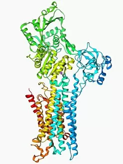
Background imageMolecular Structure Collection: Calcium ATPase ion pump F006 / 9507

Calcium ATPase ion pump F006 / 9507
Calcium ATPase ion pump, molecular model. This enzyme is found in muscle cell membranes, where it pumps calcium in and out of muscle cells and controls muscle contractions

Background imageMolecular Structure Collection: Scavenger mRNA-decapping enzyme F006 / 9505

Scavenger mRNA-decapping enzyme F006 / 9505
Scavenger mRNA-decapping enzyme, molecular model. This enzyme hydrolyses the cap that is left after 3 to 5 mRNA degradation

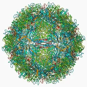
Background imageMolecular Structure Collection: Parvovirus particle, molecular model F006 / 9499

Parvovirus particle, molecular model F006 / 9499
Parvovirus particle. Molecular model showing the structure of the capsid (outer protein coat) of a human parvovirus (family Parvoviridae) particle

Background imageMolecular Structure Collection: Staphylococcal enterotoxin C2 molecule F006 / 9506

Staphylococcal enterotoxin C2 molecule F006 / 9506
Staphylococcal enterotoxin C2. Molecular model of the C2 enterotoxin from the bacterium Staphylococcus aureus

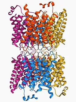
Background imageMolecular Structure Collection: Aquaporin membrane protein F006 / 9503

Aquaporin membrane protein F006 / 9503
Aquaporin membrane protein, molecular model. Aquaporins are membrane proteins that form channels (lower right) that help water molecules pass in and out of cells

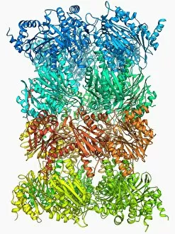
Background imageMolecular Structure Collection: Yeast enzyme, molecular model F006 / 9498

Yeast enzyme, molecular model F006 / 9498
Yeast enzyme. Molecular model of an enzyme from bakers yeast (Saccharomyces cerevisiae). This is the 20S proteasome. A proteasome is a complex type of proteinase (protein-digesting enzyme)

Background imageMolecular Structure Collection: SARS virus capsid protein F006 / 9504

SARS virus capsid protein F006 / 9504
SARS virus capsid protein, molecular model. This protein is responsible for binding the capsid (outer coat) of the SARS (severe acute respiratory syndrome)

Background imageMolecular Structure Collection: RNA-induced silencing complex F006 / 9502

RNA-induced silencing complex F006 / 9502
RNA-induced silencing complex (RISC), molecular model. This complex consists of a bacterial argonaute protein bound to a small interfering RNA (siRNA) molecule (red and blue)

Background imageMolecular Structure Collection: Photosystem II molecule F006 / 9500

Photosystem II molecule F006 / 9500
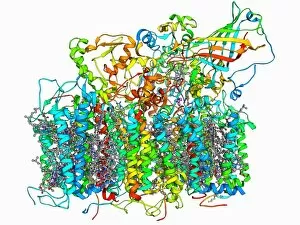
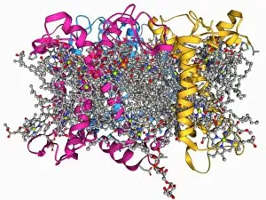
Photosystem II. Molecular model of the photosystem II complex. Photosystems are protein complexes involved in photosynthesis

Background imageMolecular Structure Collection: Photosystem II molecule F006 / 9497

Photosystem II molecule F006 / 9497
Photosystem II. Molecular model of the photosystem II complex. Photosystems are protein complexes involved in photosynthesis

Background imageMolecular Structure Collection: Nerve growth factor bound to receptor F006 / 9501

Nerve growth factor bound to receptor F006 / 9501
Nerve growth factor bound to receptor. Molecular model of nerve growth factor (NGF) bound to the p75 neurotrophin receptor. NGF is a neurotrophin that acts on the development and function of nerves

Background imageMolecular Structure Collection: Rhinovirus capsid, molecular model F006 / 9490

Rhinovirus capsid, molecular model F006 / 9490
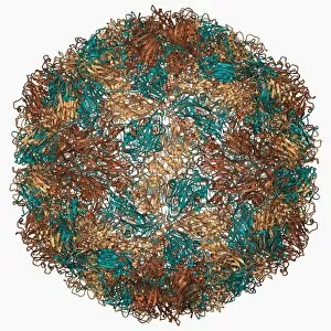
Rhinovirus capsid, molecular model. This is human rhinovirus. The rhinovirus infects the upper respiratory tract and is the cause of the common cold. It is spread by coughs and sneezes

Background imageMolecular Structure Collection: H1 antigen from 1918 influenza virus F006 / 9495

H1 antigen from 1918 influenza virus F006 / 9495
H1 antigen from 1918 influenza virus

Background imageMolecular Structure Collection: HIV reverse transcription enzyme F006 / 9494

HIV reverse transcription enzyme F006 / 9494
HIV reverse transcription enzyme. Molecular model of the reverse transcriptase enzyme (blue and green) found in HIV (the human immunodeficiency virus)

Background imageMolecular Structure Collection: EcoRV restriction enzyme molecule F006 / 9496

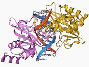
EcoRV restriction enzyme molecule F006 / 9496
EcoRV restriction enzyme. Molecular model of the type II restriction enzyme EcoRV (pink and yellow) bound to a cleaved section of DNA (deoxyribonucleic acid, red and blue)

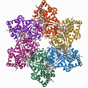
Background imageMolecular Structure Collection: Rubisco enzyme molecule F006 / 9491

Rubisco enzyme molecule F006 / 9491
Rubisco. Molecular model of the enzyme rubisco (ribulose bisphosphate carboxylase oxygenase). Rubisco is thought to be the most abundant and important protein found in nature

Background imageMolecular Structure Collection: Hammerhead ribozyme molecule F006 / 9492

Hammerhead ribozyme molecule F006 / 9492
Hammerhead ribozyme, molecular model. Ribozymes are RNA (ribonucleic acid) molecules that catalyse certain biochemical reactions

Background imageMolecular Structure Collection: Murine p97 protein molecule F006 / 9487

Murine p97 protein molecule F006 / 9487
Murine p97 protein, molecular model. This mouse enzyme, is involved in membrane fusion and ubiquitin-dependent protein degradation

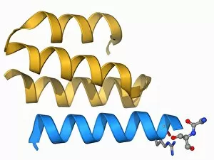
Background imageMolecular Structure Collection: Neuropeptide Y neurotransmitter molecule F006 / 9493

Neuropeptide Y neurotransmitter molecule F006 / 9493
Neuropeptide Y neurotransmitter molecule. Molecular model showing the structure of the neurotransmitter neuropeptide Y (NPY). NPY is found in the brain and autonomic nervous system

Background imageMolecular Structure Collection: Interferon gamma molecule F006 / 9489

Interferon gamma molecule F006 / 9489
Interferon gamma, molecular model. Interferon gamma is produced by certain immune cells (T cells, dendritic cells and NK cells) as part of the immune response to invading pathogens and tumours

Background imageMolecular Structure Collection: RNA interference viral suppressor and RNA F006 / 9488

RNA interference viral suppressor and RNA F006 / 9488
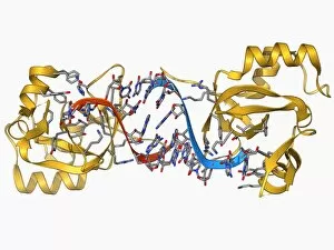
RNA interference viral suppressor and RNA. Molecular model of the p19 protein (yellow) from a Tombusvirus, suppressing a double-stranded, small interfering RNA (siRNA) molecule (red and blue)

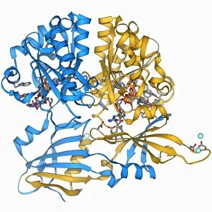
Background imageMolecular Structure Collection: Uricase enzyme, molecular model F006 / 9485

Uricase enzyme, molecular model F006 / 9485
Uricase, molecular model. This enzyme, also known as urate oxidase, catalyses the last stage of purine catabolism. It is absent from humans

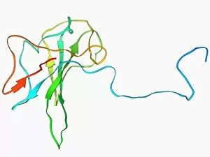
Background imageMolecular Structure Collection: Yersinia pestis virulence factor F006 / 9486

Yersinia pestis virulence factor F006 / 9486
Yersinia pestis virulence factor. Molecular model of the V-antigen protein, a virulence factor in Yersinia pestis, the causative agent of the plague

Background imageMolecular Structure Collection: Transcription factor and DNA molecule F006 / 9484

Transcription factor and DNA molecule F006 / 9484
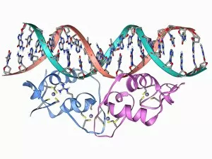
Transcription factor and DNA molecule. Molecular model of glucocorticoid receptor (GR) transcription factor protein (pink and blue) complexed with a molecule of DNA (deoxyribonucleic acid)

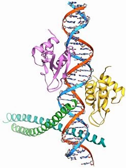
Background imageMolecular Structure Collection: Androgen receptor, molecular model F006 / 9483

Androgen receptor, molecular model F006 / 9483
Androgen receptor. Molecular model of the DNA-binding region of an androgen receptor (pink and yellow) complexed with DNA (deoxyribonucleic acid, blue and red)

Background imageMolecular Structure Collection: Human catalase, molecular model F006 / 9478

Human catalase, molecular model F006 / 9478
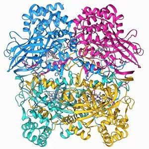
Human catalase, molecular model. This enzyme catalyses the break down of hydrogen peroxide to water and oxygen. Hydrogen peroxide is a highly toxic byproduct of a number of normal cellular processes

Background imageMolecular Structure Collection: Chinese scorpion toxin structure F006 / 9482

Chinese scorpion toxin structure F006 / 9482
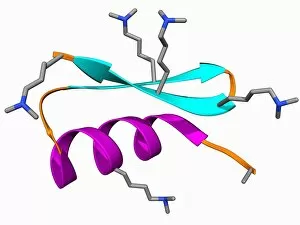
Chinese scorpion toxin structure. Molecular model of the toxin BmBKTtx1, produced by the Chinese scorpion (Buthus martensi Karsch)

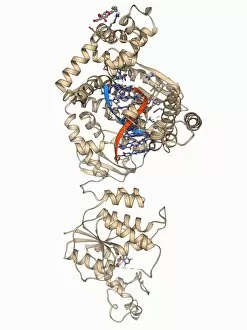
Background imageMolecular Structure Collection: Endonuclease IV molecule F006 / 9480

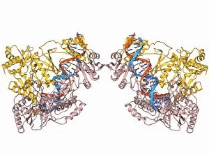
Endonuclease IV molecule F006 / 9480
Endonuclease IV molecule. Molecular model of the endonuclease IV restriction enzyme EcoRV (beige) bound to a cleaved section of DNA (deoxyribonucleic acid, blue, red and green)

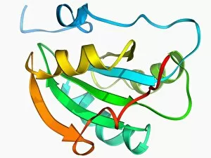
Background imageMolecular Structure Collection: Novel protein, molecular model F006 / 9481

Novel protein, molecular model F006 / 9481
Novel protein. Molecular model of a computer-designed protein named TOP7

Background imageMolecular Structure Collection: Haemagglutinin protein subunit F006 / 9479

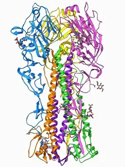
Haemagglutinin protein subunit F006 / 9479
Haemagglutinin protein subunit. Molecular model of the ectodomain of the haemagglutinin HA(2) subunit. Haemagglutinin is a surface protein from the influenza A virus

Background imageMolecular Structure Collection: Foot-and-mouth disease virus F006 / 9471

Foot-and-mouth disease virus F006 / 9471
Foot-and-mouth disease virus. Molecular model of the foot-and-mouth disease (FMD) virus Aphtae epizooticae, with antibodies (immunoglobulins)

Background imageMolecular Structure Collection: RNA polymerase molecule F006 / 9475

RNA polymerase molecule F006 / 9475
RNA polymerase. Molecular model of RNA polymerase (beige) transcribing a strand of mRNA (messenger ribonucleic acid, pink) from a DNA (deoxyribonucleic acid) template (red and blue)

Background imageMolecular Structure Collection: Cytochrome c oxidase and antibody F006 / 9474

Cytochrome c oxidase and antibody F006 / 9474
Cytochrome c oxidase. Molecular model of a cytochrome c oxidase enzyme complexed with an antibody. Cytochrome molecules perform oxidation and reduction reactions for electron transport

Background imageMolecular Structure Collection: Human prion protein, molecular model F006 / 9477

Human prion protein, molecular model F006 / 9477
Human prion protein, molecular model. Prions are abnormal proteins that cause a group of fatal neurodegenerative diseases including BSE in cows and CJD in humans

Background imageMolecular Structure Collection: Herpesvirus immune inhibitor protein F006 / 9476

Herpesvirus immune inhibitor protein F006 / 9476
Herpesvirus immune inhibitor protein. Molecular model of the protein ICP47 from the herpes simplex virus-1 (HSV-1). This protein prevents antigens from the virus being presented to immune cells

Background imageMolecular Structure Collection: Poliovirus type 3 capsid, molecular model F006 / 9465

Poliovirus type 3 capsid, molecular model F006 / 9465
Poliovirus type 3 capsid, molecular model. This enterovirus causes poliomyelitis (polio) in humans, which affects the nervous system, sometimes leading to paralysis

Background imageMolecular Structure Collection: Importin heterodimer protein F006 / 9472

Importin heterodimer protein F006 / 9472
Importin heterodimer protein. Molecular model showing the importin heterodimer, a complex between importin alpha and importin beta. Importin imports other proteins into the cell nucleus

Background imageMolecular Structure Collection: Adenovirus fibre shaft protein F006 / 9473

Adenovirus fibre shaft protein F006 / 9473
Adenovirus fibre shaft protein, molecular model. This protein forms part of the shaft of the fibres or spikes by which adenoviruses attach to receptors on cells during the infection process

Background imageMolecular Structure Collection: Haemagglutinin viral surface protein F006 / 9470

Haemagglutinin viral surface protein F006 / 9470
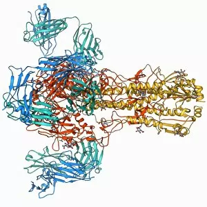
Haemagglutinin viral surface protein. Molecular model of haemagglutinin, a surface protein from the influenza virus, complexed with a neutralising antibody

Background imageMolecular Structure Collection: Outer membrane phospholipase A molecule F006 / 9469

Outer membrane phospholipase A molecule F006 / 9469
Outer membrane phospholipase A. Molecular model of the integral membrane protein, outer membrane phospholipase A from the Escherichia coli bacterium

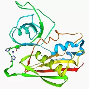
Background imageMolecular Structure Collection: Nickel-containing superoxide dismutase F006 / 9468

Nickel-containing superoxide dismutase F006 / 9468
Nickel-containing superoxide dismutase enzyme, molecular model. This enzyme scavenges and decomposes the potentially toxic first reduction product, superoxide, of aerobic respiration

Background imageMolecular Structure Collection: Lactose transporter protein molecule F006 / 9466

Lactose transporter protein molecule F006 / 9466
Lactose transporter protein. Molecular model of the transmembrane transport protein lactose permease bound with a lactose homolog

Background imageMolecular Structure Collection: Aspirin drug target molecule F006 / 9464

Aspirin drug target molecule F006 / 9464
Aspirin drug target. Molecular model of the enzyme prostaglandin H2 synthase (PGHS), the target of the anti-inflammatory drug aspirin


All Professionally Made to Order for Quick Shipping

"Molecular Structure: Unlocking the Secrets of Life's Building Blocks" From anaesthetics inhibiting ion channels to antidepressant molecules, the intricate world holds endless wonders. The C015 / 6718 anaesthetic molecule delicately interacts with ion channels, altering their function and providing relief from pain. Meanwhile, Amitriptyline, an antidepressant molecule, works its magic by modulating neurotransmitters in our brains. In the realm of immunity, Immunoglobulin G antibody F007 / 9894 stands tall as a defender against pathogens. Its unique structure allows it to recognize and neutralize foreign invaders effectively. On another front, DNA artwork showcases the elegance and complexity that underlies all life forms on Earth. Creatine amino acid molecule fuels our muscles during intense physical activities while nanotube technology revolutionizes various industries with its exceptional properties. These tiny tubes hold immense potential for advancements in medicine and materials science alike. Zinc fingers bound to a DNA strand demonstrate how proteins can precisely interact with genetic material. This interaction plays a crucial role in gene regulation and expression. Carbon nanotubes take center stage once again as they exhibit remarkable strength and conductivity at the nano-scale level. Oxytocin neurotransmitter molecule reminds us of love's powerful influence on human connections—its presence promotes bonding between individuals. Manganese superoxide dismutase enzyme F006 / 9423 safeguards our cells by combating harmful free radicals that contribute to aging and disease. Even viruses have their own molecular structures; SARS coronavirus protein represents one such example—a key player in viral replication within host cells. Conceptual artwork further explores nanotube technology's limitless possibilities—the fusion of imagination and scientific innovation knows no bounds here. As we delve deeper into understanding molecular structures, we unravel nature's blueprint for life itself—one atom at a time. These captivating glimpses into the microscopic world remind us of both the fragility and resilience found within the building blocks of existence.