Fraud Blocker Skip to main content

Molecular Structure Collection (page 16)

"Molecular Structure: Unlocking the Secrets of Life's Building Blocks" From anaesthetics inhibiting ion channels to antidepressant molecules

Background imageMolecular Structure Collection: Integrin transmembrane domain F006 / 9614

Integrin transmembrane domain F006 / 9614
Integrin transmembrane domain, molecular model. Integrins are transmembrane cell adhesion receptors

Background imageMolecular Structure Collection: Cytochrome P450 and erythromycin F006 / 9610

Cytochrome P450 and erythromycin F006 / 9610
Cytochrome P450 and erythromycin. Molecular model of human cytochrome P450 complexed with the antibiotic erythromycin. This protein plays a crucial role in metabolism in animals (including humans)

Background imageMolecular Structure Collection: Growth factor receptor molecule F006 / 9613

Growth factor receptor molecule F006 / 9613
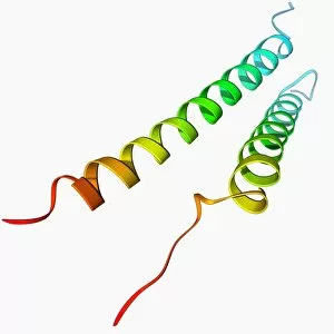
Growth factor receptor. Molecular model of the transmembrane segment of the ErbB2 growth factor receptor

Background imageMolecular Structure Collection: Nerve growth factor bound to receptor F006 / 9608

Nerve growth factor bound to receptor F006 / 9608
Nerve growth factor. Molecular model of human nerve growth factor bound to the TrkA receptor. NGF is a neurotrophin that acts on the development and function of nerves

Background imageMolecular Structure Collection: Viral RNA packaging signal complex F006 / 9609

Viral RNA packaging signal complex F006 / 9609
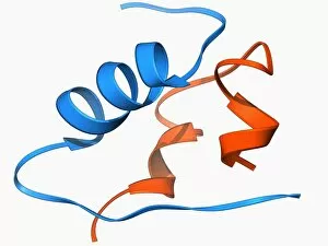
Viral RNA packaging signal complex. Molecular model of the muPsi RNA packaging signal complex from the Rous sarcoma vuris

Background imageMolecular Structure Collection: HIV reverse transcription enzyme F006 / 9606

HIV reverse transcription enzyme F006 / 9606
HIV reverse transcription enzyme. Molecular model of the reverse transcriptase enzyme (pink) found in HIV (the human immunodeficiency virus)

Background imageMolecular Structure Collection: Haemoglobin, molecular model F006 / 9604

Haemoglobin, molecular model F006 / 9604
Haemoglobin, molecular model. This is deoxyhaemoglobin, the molecule in its non-oxygen bound state. Haemoglobin transports oxygen around the body in red blood cells
Background imageMolecular Structure Collection: Rhomboid protease molecule F006 / 9607

Rhomboid protease molecule F006 / 9607
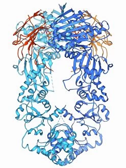
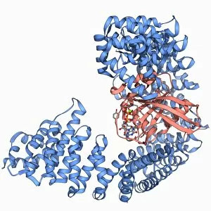
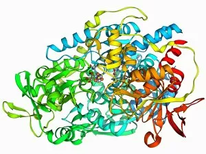
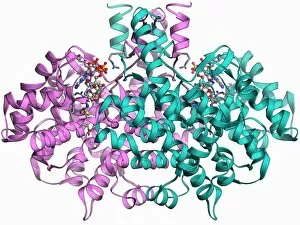
Rhomboid protease. Molecular model of the rhomboid protease enzyme GlpG from the bacterium Escherichia coli. Proteases are enzymes that break down proteins

Background imageMolecular Structure Collection: Glutamine synthetase enzyme F006 / 9598

Glutamine synthetase enzyme F006 / 9598
Glutamine synthetase enzyme, molecular model. This ligase enzyme forms chemical bonds between molecules. It plays an important role in the metabolism of nitrogen by catalysing the condensation of

Background imageMolecular Structure Collection: Insulin molecule F006 / 9605

Insulin molecule F006 / 9605
Insulin molecule. Molecular model of the hormone insulin. Insulin consists of two peptide chains, A and B, which are linked by disulphide bridges

Background imageMolecular Structure Collection: 3-hydroxyacyl-CoA dehydrogenase F006 / 9602

3-hydroxyacyl-CoA dehydrogenase F006 / 9602
3-hydroxyacyl-CoA dehydrogenase, molecular model. This enzyme is found in human heart tissue, and catalyses a reaction that is part of the beta-oxidation pathway

Background imageMolecular Structure Collection: Hepatitis B virus capsid, molecular model F006 / 9594

Hepatitis B virus capsid, molecular model F006 / 9594
Hepatitis B virus capsid, molecular model. This virus, transmitted through infected bodily fluids or blood, causes the disease hepatitis B in humans, leading to acute liver inflammation

Background imageMolecular Structure Collection: Thrombin protein, molecular model F006 / 9603

Thrombin protein, molecular model F006 / 9603
Thrombin protein, molecular model. Thrombin is an enzyme involved in the blood coagulation (clotting) process. It converts fibrinogen (a soluble plasma glycoprotein synthesised in the liver)

Background imageMolecular Structure Collection: Haemoglobin S, molecular model F006 / 9601

Haemoglobin S, molecular model F006 / 9601
Haemoglobin S. Molecular model of the mutant form of haemoglobin (haemoglobin S) that causes sickle cell anaemia. This is deoxyhaemoglobin S, the molecule in its non-oxygen bound state

Background imageMolecular Structure Collection: RNA-editing enzyme, molecular model F006 / 9599

RNA-editing enzyme, molecular model F006 / 9599
RNA-editing enzyme. Molecular model of a left-handed, RNA double helix (Z-RNA, centre) bound by the Z alpha domain of the human RNA-editing enzyme ADAR1 (double-stranded RNA adenosine deaminase)

Background imageMolecular Structure Collection: Protein kinase regulatory subunit F006 / 9600

Protein kinase regulatory subunit F006 / 9600
Protein kinase regulatory subunit. Molecular model of a regulatory subunit from cAMP-dependent protein kinase bound to. This enzyme is also known as protein kinase A (PKA)

Background imageMolecular Structure Collection: Cyanobacterial circadian clock protein F006 / 9595

Cyanobacterial circadian clock protein F006 / 9595
Cyanobacterial circadian clock protein, molecular model. This protein is a kinase known as KaiC. Its structure is a hexamer

Background imageMolecular Structure Collection: Programmed cell death protein molecule F006 / 9597

Programmed cell death protein molecule F006 / 9597
Human programmed cell death protein 4, molecular model. This protein is involved in apoptosis (programmed cell death)

Background imageMolecular Structure Collection: Multidrug transporter molecule F006 / 9596

Multidrug transporter molecule F006 / 9596
Multidrug transporter. Molecular model of the multidrug transporter EmrD from the bacterium Escherichia coli. This protein pumps drugs, including antibiotics, out of the bacterial cell

Background imageMolecular Structure Collection: Respiratory complex I F006 / 9593

Respiratory complex I F006 / 9593
Respiratory complex I. Molecular model of the hydrophilic domain of respiratory complex I from the bacterium Thermus thermophilus. This enzyme is involved in energy production

Background imageMolecular Structure Collection: Chymotrypsin digestive enzyme molecule F006 / 9577

Chymotrypsin digestive enzyme molecule F006 / 9577
Chymotrypsin digestive enzyme, molecular model. Chymotrypsin is a protease, an enzyme that breaks down proteins and peptides. It is secreted into the duodenum (small intestine) by the pancreas

Background imageMolecular Structure Collection: Ribonuclease bound to transfer RNA F006 / 9591

Ribonuclease bound to transfer RNA F006 / 9591
Ribonuclease bound to transfer RNA, molecular model. This complex consists of the ribonuclease Z (RNase Z, green and pink) enzyme bound to a transfer RNA (tRNA) molecule (orange and blue)

Background imageMolecular Structure Collection: H5N1 Haemagglutinin protein subunit F006 / 9590

H5N1 Haemagglutinin protein subunit F006 / 9590
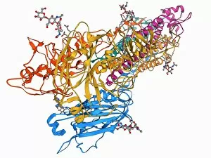
H5N1 haemagglutinin protein subunit. Molecular model of the haemagglutinin HA(5) subunit. Haemagglutinin is a surface protein from the influenza A virus

Background imageMolecular Structure Collection: RNA-induced silencing complex F006 / 9587

RNA-induced silencing complex F006 / 9587
RNA-induced silencing complex (RISC), molecular model. This complex consists of a bacterial argonaute protein (top right) bound to a small interfering RNA (siRNA) molecule (red and blue)

Background imageMolecular Structure Collection: RNA interference protein, molecular model F006 / 9589

RNA interference protein, molecular model F006 / 9589
RNA interference protein, molecular model. This RNA interference protein is also known as dicer. It is an RNAase enzyme that cleaves double-stranded RNA into short fragments called small interfering

Background imageMolecular Structure Collection: Acyl carrier protein molecule F006 / 9588

Acyl carrier protein molecule F006 / 9588
Acyl carrier protein (ACP), molecular model. This enzyme is involved in fatty acid synthesis

Background imageMolecular Structure Collection: DNA transcription, molecular model F006 / 9584

DNA transcription, molecular model F006 / 9584
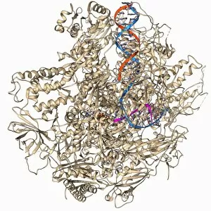
DNA transcription. Molecular model of the enzyme RNA polymerase II synthesising a mRNA (messenger ribonucleic acid) strand from a DNA (deoxyribonucleic acid) template

Background imageMolecular Structure Collection: Dynamin enzyme, molecular model F006 / 9583

Dynamin enzyme, molecular model F006 / 9583
Dynamin enzyme. Molecular model of the pleckstrin homology (PH) domain of the dynamin enzyme. Domains are structural regions of enzymes that are often actively involved in biological processes

Background imageMolecular Structure Collection: Double-stranded RNA-ribonuclease III F006 / 9585

Double-stranded RNA-ribonuclease III F006 / 9585
Double-stranded RNA-ribonuclease III. Molecular model of ribonuclease III (RNase III, D44N, pink and green) complexed with a double-stranded RNA (ribonucleic acid) strand (red and blue)

Background imageMolecular Structure Collection: Human rotavirus capsid protein molecule F006 / 9582

Human rotavirus capsid protein molecule F006 / 9582
Human rotavirus capsid protein, molecular model

Background imageMolecular Structure Collection: Insulin receptor molecule F006 / 9581

Insulin receptor molecule F006 / 9581
Insulin receptor, molecular model. The insulin receptor is a transmembrane protein, that is it spans the cellular membrane

Background imageMolecular Structure Collection: Haemoglobin, molecular model F006 / 9580

Haemoglobin, molecular model F006 / 9580
Haemoglobin, molecular model. This is deoxyhaemoglobin, the molecule in its non-oxygen bound state. Haemoglobin transports oxygen around the body in red blood cells

Background imageMolecular Structure Collection: Bioluminescent enzyme molecule F006 / 9579

Bioluminescent enzyme molecule F006 / 9579
Bioluminescent enzyme. Molecular model of the bioluminescent enzyme luciferase from the Japanese aquatic firefly (Luciola cruciata) complexed with an intermediate protein

Background imageMolecular Structure Collection: Adenovirus penton base protein F006 / 9572

Adenovirus penton base protein F006 / 9572
Adenovirus penton base protein, molecular model. This protein molecule is a subunit called a penton, forming the vertices of the capsid of this adenovirus

Background imageMolecular Structure Collection: Fatty acid synthase molecule F006 / 9575

Fatty acid synthase molecule F006 / 9575
Fatty acid synthase (FAS), molecular model. FAS is a multi-enzyme that plays a key role in the synthesis of fatty acids (lipids) in the human body. It is not a single enzyme but a whole enzyme system

Background imageMolecular Structure Collection: Heat shock protein 90 chaperone complex F006 / 9576

Heat shock protein 90 chaperone complex F006 / 9576
Heat shock protein 90 (Hsp90) chaperone complex, molecular model. Hsp90 is a molecular chaperone and a member of the heat shock protein (HSP) family

Background imageMolecular Structure Collection: Chymotrypsin inhibitor 2 molecule F006 / 9578

Chymotrypsin inhibitor 2 molecule F006 / 9578
Chymotrypsin inhibitor 2, molecular model. This enzyme is a serine protease inhibitor (serpin) found in barley (Hordeum vulgare) seeds

Background imageMolecular Structure Collection: Citrate synthase molecule F006 / 9573

Citrate synthase molecule F006 / 9573
Citrate synthase, molecular model. This enzyme is involved in the first step of the citric acid (or Krebs) cycle, the process by which mitochondria convert glucose to energy

Background imageMolecular Structure Collection: Family 84 glycoside hydrolase molecule F006 / 9574

Family 84 glycoside hydrolase molecule F006 / 9574
Family 84 glycoside hydrolase, molecular model. Glycoside hydrolases are enzymes that hydrolase the glycosidic bond between two or more carbohydrates

Background imageMolecular Structure Collection: Acetylcholine receptor molecule F006 / 9569

Acetylcholine receptor molecule F006 / 9569
Acetylcholine receptor. Molecular model showing the structure of a nicotinic acetlycholine receptor. This receptor, for the neurotransmitter acetylcholine

Background imageMolecular Structure Collection: Nuclear import complex molecule F006 / 9571

Nuclear import complex molecule F006 / 9571
Nuclear import complex. Molecular model of an importin protein complexed with a RanGTP molecule. This protein complex imports other proteins into the cell nucleus

Background imageMolecular Structure Collection: Retinal-producing oxygenase enzyme F006 / 9570

Retinal-producing oxygenase enzyme F006 / 9570
Retinal-producing oxygenase enzyme, molecular model. This enzyme plays a role in the production and metabolism of retinal and other apocarotenoids. Retinal is the chemical basis of vision in animals

Background imageMolecular Structure Collection: Flu virus surface protein molecule F006 / 9566

Flu virus surface protein molecule F006 / 9566
Flu virus surface protein molecule. Molecular model of the neuraminidase glycoprotein enzyme found on the surface of the influenza A (flu) virus

Background imageMolecular Structure Collection: Amyloid beta protein molecule F006 / 9568

Amyloid beta protein molecule F006 / 9568
Amyloid beta protein. Molecular model showing the structure of amyloid beta protein, the primary component of amyloid plaques found in the brains of Alzheimers patients

Background imageMolecular Structure Collection: Calcium-binding protein molecule F006 / 9567

Calcium-binding protein molecule F006 / 9567
Calcium-binding protein. Molecule model of the calcium-binding protein calmodulin (CaM) bound to a myosin light-chain kinase molecule. This complex is involved in muscle contraction

Background imageMolecular Structure Collection: Iron-regulatory protein, molecular model F006 / 9565

Iron-regulatory protein, molecular model F006 / 9565
Iron-regulatory protein. Molecular model of iron regulatory protein 1 (IRP1). Depending on the conformation of IRP1, it can acts as either a regulator of mRNA (messenger RNA) or an enzyme

Background imageMolecular Structure Collection: Voltage-gated potassium channel F006 / 9562

Voltage-gated potassium channel F006 / 9562
Voltage-gated potassium (Kv) ion channel, molecular model. Ion channels are membrane-spanning proteins that form pores in cell membranes

Background imageMolecular Structure Collection: Tumour suppressor protein with DNA F006 / 9564

Tumour suppressor protein with DNA F006 / 9564
Tumour suppressor protein. Molecular model of the tumour suppressor protein p53 (beige) bound to a molecule of DNA (deoxyribonucleic acid, red and blue)


All Professionally Made to Order for Quick Shipping

"Molecular Structure: Unlocking the Secrets of Life's Building Blocks" From anaesthetics inhibiting ion channels to antidepressant molecules, the intricate world holds endless wonders. The C015 / 6718 anaesthetic molecule delicately interacts with ion channels, altering their function and providing relief from pain. Meanwhile, Amitriptyline, an antidepressant molecule, works its magic by modulating neurotransmitters in our brains. In the realm of immunity, Immunoglobulin G antibody F007 / 9894 stands tall as a defender against pathogens. Its unique structure allows it to recognize and neutralize foreign invaders effectively. On another front, DNA artwork showcases the elegance and complexity that underlies all life forms on Earth. Creatine amino acid molecule fuels our muscles during intense physical activities while nanotube technology revolutionizes various industries with its exceptional properties. These tiny tubes hold immense potential for advancements in medicine and materials science alike. Zinc fingers bound to a DNA strand demonstrate how proteins can precisely interact with genetic material. This interaction plays a crucial role in gene regulation and expression. Carbon nanotubes take center stage once again as they exhibit remarkable strength and conductivity at the nano-scale level. Oxytocin neurotransmitter molecule reminds us of love's powerful influence on human connections—its presence promotes bonding between individuals. Manganese superoxide dismutase enzyme F006 / 9423 safeguards our cells by combating harmful free radicals that contribute to aging and disease. Even viruses have their own molecular structures; SARS coronavirus protein represents one such example—a key player in viral replication within host cells. Conceptual artwork further explores nanotube technology's limitless possibilities—the fusion of imagination and scientific innovation knows no bounds here. As we delve deeper into understanding molecular structures, we unravel nature's blueprint for life itself—one atom at a time. These captivating glimpses into the microscopic world remind us of both the fragility and resilience found within the building blocks of existence.