Fraud Blocker Skip to main content

Molecular Structure Collection (page 21)

"Molecular Structure: Unlocking the Secrets of Life's Building Blocks" From anaesthetics inhibiting ion channels to antidepressant molecules

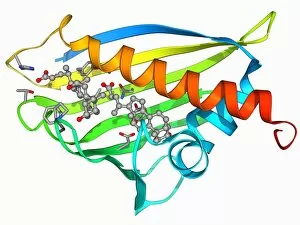
Background imageMolecular Structure Collection: Cholesterol producing enzyme molecule F006 / 9365

Cholesterol producing enzyme molecule F006 / 9365
Cholesterol producing enzyme. Molecular model of HMG-CoA reductase (HMGCR), the rate-controlling enzyme of the metabolic pathway that produces cholesterol in the body

Background imageMolecular Structure Collection: Bacterial cell wall enzyme molecule F006 / 9364

Bacterial cell wall enzyme molecule F006 / 9364
Bacterial cell wall enzyme. Molecular model of D-alanyl-D-alanine carboxypeptidase, a transpeptidase. This enzyme cross-links peptidoglycan chains in bacterial cell walls, making them rigid

Background imageMolecular Structure Collection: Mitochondrial processing peptidase F006 / 9362

Mitochondrial processing peptidase F006 / 9362
Yeast mitochondrial processing peptidase, molecular model. This enzyme plays an essential role in importing mitochondrial proteins

Background imageMolecular Structure Collection: MHC protein-antigen complex F006 / 9363

MHC protein-antigen complex F006 / 9363
MHC protein-antigen complex. Molecular model of the human class I MHC (major histocompatibility complex) protein HLA-B27 complexed with beta-2 microglobulin

Background imageMolecular Structure Collection: Phosphoglucose isomerase molecule F006 / 9361

Phosphoglucose isomerase molecule F006 / 9361
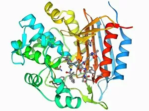
Phosphoglucose isomerase molecule. Molecular model of a rabbit phosphoglucose isomerase molecule complexed with a fructose-6-phosphate molecule

Background imageMolecular Structure Collection: HIV reverse transcription enzyme F006 / 9360

HIV reverse transcription enzyme F006 / 9360
HIV reverse transcription enzyme. Molecular model of the reverse transcriptase enzyme found in HIV (the human immunodeficiency virus)

Background imageMolecular Structure Collection: HGPRTase molecule F006 / 9359

HGPRTase molecule F006 / 9359
HGPRTase. Molecular model of hypoxanthine-guanine phosphoribosyltransferase (HGPRTase) bound to a molecule of guanosine monophosphate (GMP). HGPRTase is involved in the purine salvage pathway

Background imageMolecular Structure Collection: Haemoglobin molecule F006 / 9356

Haemoglobin molecule F006 / 9356
Haemoglobin, molecular model. Haemoglobin is a metalloprotein that transports oxygen around the body in red blood cells. Each molecule consists of iron-containing haem groups (sticks)

Background imageMolecular Structure Collection: Beta-actin molecule F006 / 9358

Beta-actin molecule F006 / 9358
Beta-actin. Molecular model of the cytoskeletal protein beta-actin bound to profilin. The cytoskeleton maintains the cells shape

Background imageMolecular Structure Collection: Human prion precursor protein F006 / 9357

Human prion precursor protein F006 / 9357
Human prion precursor protein, molecular model showing secondary structure. The human prion protein (hPrP) is a prion precursor

Background imageMolecular Structure Collection: Serum transferrin molecule F006 / 9353

Serum transferrin molecule F006 / 9353
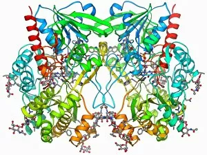
Serum transferrin, molecular model. Transferrins are iron-binding glycoproteins found in blood plasma

Background imageMolecular Structure Collection: Oestrogen receptor bound to DNA F006 / 9354

Oestrogen receptor bound to DNA F006 / 9354
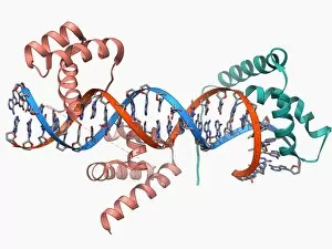
Oestrogen receptor bound to DNA. Molecular model of the DNA-binding domain (green and pink) of the oestrogen receptor bound to a molecule of DNA (deoxyribonucleic acid, blue and orange)

Background imageMolecular Structure Collection: Human growth hormone molecule F006 / 9355

Human growth hormone molecule F006 / 9355
Human growth hormone (hGH), molecular model. hGH is produced in the anterior pituitary gland of the brain. It binds to specific receptors on cells in the body to stimulate growth

Background imageMolecular Structure Collection: Ebola matrix protein molecule F006 / 9352

Ebola matrix protein molecule F006 / 9352
Ebola matrix protein. Molecular model of the Ebola virus matrix protein VP40 bound to RNA (ribonucleic acid). This membrane-associated protein is thought to be necessary for the assembly

Background imageMolecular Structure Collection: Haemoglobin molecule F006 / 9350

Haemoglobin molecule F006 / 9350
Haemoglobin, molecular model. Haemoglobin is a metalloprotein that transports oxygen around the body in red blood cells. Each molecule consists of iron-containing haem groups (sticks)

Background imageMolecular Structure Collection: Glycogen phosphorylase molecule F006 / 9347

Glycogen phosphorylase molecule F006 / 9347
Glycogen phosphorylase, molecular model. This is an enzyme involved in breaking down glycogen, the energy storage molecule involved in animal metabolism

Background imageMolecular Structure Collection: Glucose oxidase molecule F006 / 9348

Glucose oxidase molecule F006 / 9348
Glucose oxidase, molecular model. In cells this enzyme catalyses the breakdown of glucose into its metabolites

Background imageMolecular Structure Collection: Insulin-like growth 1 factor molecule F006 / 9351

Insulin-like growth 1 factor molecule F006 / 9351
Insulin-like growth factor 1 (IGF-1), molecular model. IGFs are polypeptides that are similar in their molecular structure to insulin

Background imageMolecular Structure Collection: Ribozyme enzyme and RNA F006 / 9346

Ribozyme enzyme and RNA F006 / 9346
Ribozyme enzyme and RNA, molecular model. Ribozymes are RNA (ribonucleic acid) molecules that catalyse certain biochemical reactions

Background imageMolecular Structure Collection: Transcription factors bound to DNA F006 / 9349

Transcription factors bound to DNA F006 / 9349
Transcription factors bound to DNA. Molecular model of the Oct4 (pink) and Sox2 (green) transcription factors bound to a molecule of DNA (deoxyribonucleic acid, red and blue)

Background imageMolecular Structure Collection: GA bacteriophage capsid F006 / 9341

GA bacteriophage capsid F006 / 9341
GA bacteriophage capsid, molecular model. Bacteriophages are viruses that infect bacteria, with the capsid forming the bacteriophage head

Background imageMolecular Structure Collection: Heterotrimeric G protein complex molecule F006 / 9344

Heterotrimeric G protein complex molecule F006 / 9344
Heterotrimeric G protein complex molecule. Molecular model of a heterotrimeric G protein complex. This protein has three different subunits (heterotrimer), alpha, beta and gamma

Background imageMolecular Structure Collection: SV40 antigen and tumor suppressor F006 / 9345

SV40 antigen and tumor suppressor F006 / 9345
SV40 antigen and tumour suppressor. Molecular model of the simian virus (SV40) large T antigen bound to a retinoblastoma tumour suppressor (Rb)

Background imageMolecular Structure Collection: Bacterial protease molecule F006 / 9340

Bacterial protease molecule F006 / 9340
Bacterial protease molecule. Computer model of a molecule of HsIUV protease complexed with its chaperone protein from a bacterium. Proteases are enzymes that break down proteins

Background imageMolecular Structure Collection: Glutamine synthetase enzyme F006 / 9338

Glutamine synthetase enzyme F006 / 9338
Glutamine synthetase enzyme, molecular model. This ligase enzyme forms chemical bonds between molecules. It plays an important role in the metabolism of nitrogen by catalysing the condensation of

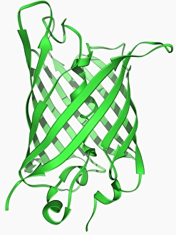
Background imageMolecular Structure Collection: Green fluorescent protein molecule F006 / 9343

Green fluorescent protein molecule F006 / 9343
Green fluorescent protein (GFP), molecular model. The molecule has a cylindrical structure formed from beta sheets (ribbons). GFP is found in the Pacific jellyfish Aequorea victoria

Background imageMolecular Structure Collection: Valyl-tRNA synthetase molecule F006 / 9342

Valyl-tRNA synthetase molecule F006 / 9342
Valyl-tRNA synthetase protein molecule. Molecular model showing bacterial valyl-tRNA synthetase complexed with valyl tRNA (transfer ribonucleic acid)

Background imageMolecular Structure Collection: Xanthine dehydrogenase molecule F006 / 9336

Xanthine dehydrogenase molecule F006 / 9336
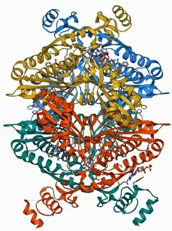
Xanthine dehydrogenase molecule. Molecular model of the Xanthine dehydrogenase (XDH) enzyme. XDH is an oxidoreductase enzyme that catalyses the last two steps in the formation of urate (uric acid)

Background imageMolecular Structure Collection: T cell receptor antigen complex F006 / 9339

T cell receptor antigen complex F006 / 9339
T cell receptor antigen complex. Molecular model of the alphabeta T cell receptor bound to the influenza haemagglutinin antigen and MHC class II molecule HLA-DR1

Background imageMolecular Structure Collection: Transcription factor complexed with DNA F006 / 9337

Transcription factor complexed with DNA F006 / 9337
Transcription factor complexed with DNA. Molecular model showing the transcriptional factor c-fos c-jun (yellow and pink) bound to a strand of DNA (deoxyribonucleic acid, orange and blue)

Background imageMolecular Structure Collection: DNA Holliday junction complex F006 / 9334

DNA Holliday junction complex F006 / 9334
DNA Holliday junction complex. Molecular model of the enzyme FLP recombinase in complex with a Holliday junction between homologous strands of DNA (deoxyribonucleic acid)

Background imageMolecular Structure Collection: Birch pollen allergen F006 / 9335

Birch pollen allergen F006 / 9335
Birch pollen allergen. Molecular model of Bet v 1l, the molecule responsible for allergic reactions to birch pollen. Here

Background imageMolecular Structure Collection: Iron storage molecule F006 / 9331

Iron storage molecule F006 / 9331
Iron storage molecule. Molecular model of ferritin, a protein that acts as an iron store and is mainly found in the liver, kidneys and spleen

Background imageMolecular Structure Collection: Bacterial ribosome, molecular model F006 / 9332

Bacterial ribosome, molecular model F006 / 9332
Bacterial ribosome. Molecular model of a 30S (small) ribosomal sub-unit from the bacteria Thermus thermophilus. Ribosomes are composed of protein and RNA (ribonucleic acid)

Background imageMolecular Structure Collection: Beta secretase enzyme, molecular model F006 / 9333

Beta secretase enzyme, molecular model F006 / 9333
Beta secretase enzyme. Molecular model of the enzyme beta secretase bound to an inhibitor molecule. Beta secretase is a membrane-associated aspartic protease

Background imageMolecular Structure Collection: Archaeon ribosome, molecular model F006 / 9328

Archaeon ribosome, molecular model F006 / 9328
Archaeon ribosome. Molecular model showing the structure of a 50S (large) ribosome from the archaeon Haloarcula marismortui. Ribosomes are composed of protein and RNA (ribonucleic acid)

Background imageMolecular Structure Collection: Isoleucyl-tRNA synthetase molecule F006 / 9329

Isoleucyl-tRNA synthetase molecule F006 / 9329
Isoleucyl-tRNA synthetase protein molecule. Molecular model showing bacterial isoleucyl-tRNA synthetase complexed with aspartyl tRNA (transfer ribonucleic acid)

Background imageMolecular Structure Collection: Interferon gamma molecule and receptor F006 / 9330

Interferon gamma molecule and receptor F006 / 9330
Interferon gamma. Molecular model of the interferon gamma dimer attached to the alpha chain of the interferon gamma receptor

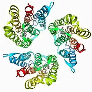
Background imageMolecular Structure Collection: Bacteriorhodopsin protein F006 / 9327

Bacteriorhodopsin protein F006 / 9327
Bacteriorhodopsin protein. Molecular model showing the structure of bacteriorhodopsin (bR), a protein found in primitive micro-organisms known as Archaea. This protein acts as a proton pump

Background imageMolecular Structure Collection: Tetraubiquitin protein molecule F006 / 9326

Tetraubiquitin protein molecule F006 / 9326
Tetraubiquitin protein, molecular model. Ubiquitin is found in all eukaryotic cells. When a protein is damaged or old it will be tagged by several ubiquitin molecules

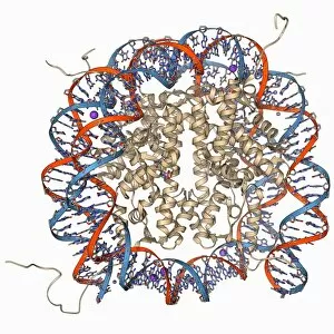
Background imageMolecular Structure Collection: Nucleosome molecule F006 / 9323

Nucleosome molecule F006 / 9323
Nucleosome, molecular model. A nucleosome is a subunit of chromatin, the substance that forms chromosomes. It consists of a short length of DNA (deoxyribonucleic acid)

Background imageMolecular Structure Collection: Rhodopsin molecule F006 / 9325

Rhodopsin molecule F006 / 9325
Rhodopsin molecule. Molecular model of the rhodopsin complex. Rhodopsin (also called visual purple) is a pigment found in the rod photoreceptor cells in the retina of the eye

Background imageMolecular Structure Collection: Cre-Lox recombination, molecular model F006 / 9322

Cre-Lox recombination, molecular model F006 / 9322
Cre-Lox recombination. Molecular model of the enzyme CRE (cyclization recombination) recombinase (beige) mediating the recombination of strands of DNA (deoxyribonucleic acid, red and blue)

Background imageMolecular Structure Collection: Proteinase inhibitor molecule F006 / 9321

Proteinase inhibitor molecule F006 / 9321
Proteinase inhibitor. Molecular model of a proteinase inhibitor, or antitrypsin, molecule bound to a tryspin protease. The proteinase inhibitor is a type of serine protease inhibitor (serpin)

Background imageMolecular Structure Collection: Voltage-gated potassium channel F006 / 9324

Voltage-gated potassium channel F006 / 9324
Voltage-gated potassium channel. Molecular model of a voltage-gated potassium (Kv) ion channel. Ion channels are membrane-spanning proteins that form pores in cell membranes

Background imageMolecular Structure Collection: Insect antifreeze protein F006 / 9320

Insect antifreeze protein F006 / 9320
Insect antifreeze protein. Molecular model of an insect antifreeze protein (AFP) from the Tenebrio molitor beetle

Background imageMolecular Structure Collection: Nucleosome molecule F006 / 9314

Nucleosome molecule F006 / 9314
Nucleosome, molecular model. A nucleosome is a subunit of chromatin, the substance that forms chromosomes. It consists of a short length of DNA (deoxyribonucleic acid)

Background imageMolecular Structure Collection: Ebola matrix protein molecule F006 / 9317

Ebola matrix protein molecule F006 / 9317
Ebola matrix protein. Molecular model of the Ebola virus matrix protein VP40. This membrane-associated protein is thought to be necessary for the assembly and budding of viral particles


All Professionally Made to Order for Quick Shipping

"Molecular Structure: Unlocking the Secrets of Life's Building Blocks" From anaesthetics inhibiting ion channels to antidepressant molecules, the intricate world holds endless wonders. The C015 / 6718 anaesthetic molecule delicately interacts with ion channels, altering their function and providing relief from pain. Meanwhile, Amitriptyline, an antidepressant molecule, works its magic by modulating neurotransmitters in our brains. In the realm of immunity, Immunoglobulin G antibody F007 / 9894 stands tall as a defender against pathogens. Its unique structure allows it to recognize and neutralize foreign invaders effectively. On another front, DNA artwork showcases the elegance and complexity that underlies all life forms on Earth. Creatine amino acid molecule fuels our muscles during intense physical activities while nanotube technology revolutionizes various industries with its exceptional properties. These tiny tubes hold immense potential for advancements in medicine and materials science alike. Zinc fingers bound to a DNA strand demonstrate how proteins can precisely interact with genetic material. This interaction plays a crucial role in gene regulation and expression. Carbon nanotubes take center stage once again as they exhibit remarkable strength and conductivity at the nano-scale level. Oxytocin neurotransmitter molecule reminds us of love's powerful influence on human connections—its presence promotes bonding between individuals. Manganese superoxide dismutase enzyme F006 / 9423 safeguards our cells by combating harmful free radicals that contribute to aging and disease. Even viruses have their own molecular structures; SARS coronavirus protein represents one such example—a key player in viral replication within host cells. Conceptual artwork further explores nanotube technology's limitless possibilities—the fusion of imagination and scientific innovation knows no bounds here. As we delve deeper into understanding molecular structures, we unravel nature's blueprint for life itself—one atom at a time. These captivating glimpses into the microscopic world remind us of both the fragility and resilience found within the building blocks of existence.