Framed Print > Popular Themes > Human Body
Framed Print : Paprosky femur defect classification C016 / 6621


![]()
Framed Photos from Science Photo Library
Paprosky femur defect classification C016 / 6621
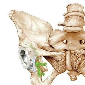
Paprosky femur defect classification. Diagram showing the classification system for femur cortex defects used to assess revision (replacement or repair) of a hip implant. The ball and socket part of the implant is at top. The rest of the implant consists of a shaft inside the femur (thigh bone). The amount of degradation in the cortical (outer) bone layer determines the amount of bone grafting needed, and whether a cementless implant can replace a cemented one. Six regions are labelled, and five subtypes described (type IV not shown). Named for US surgeon Wayne G. Paprosky, the classification was developed in the 1980s and 1990s. For specific types, see images C016/6622 to C016/6637
Science Photo Library features Science and Medical images including photos and illustrations
Media ID 9214347
© D & L GRAPHICS / SCIENCE PHOTO LIBRARY
Arthrology Arthroplasty Bone Cement Cemented Cortex Cortical Defect Defects Diagram Femur Grafting Hip Implant Hip Replacement Hip Revision Joint Label Labelled Labels Lateral Medial Orthopaedic Orthopaedics Orthopedic Orthopedics Osteological Osteology Prostheses Prosthesis Prosthetic Prosthetics Proximal Repair Replacement Surgery Surgical Type Type 1 Type 2 Type Ii Types Condition Cutouts Disorder Type 3
18"x14" Modern Frame
Introducing the Media Storehouse Framed Prints featuring the intriguing "Paprosky Femur Defect Classification C016 / 6621" image by D & L Graphics / Science Photo Library. This captivating print showcases the comprehensive Paprosky classification system for assessing femur cortex defects during hip implant revisions. Ideal for medical professionals, students, or anyone with an interest in orthopedics, this framed print adds a touch of intellectual sophistication to any workspace or home decor. The high-quality print is beautifully framed and ready to hang, ensuring a lasting impression.
16x12 Print in an MDF Wooden Frame with 180 gsm Satin Finish Paper. Glazed using shatter proof thin plexiglass. Frame thickness is 1 inch and depth 0.75 inch. Fluted cardboard backing held with clips. Supplied ready to hang with sawtooth hanger and rubber bumpers. Spot clean with a damp cloth. Packaged foam wrapped in a card.
Contemporary Framed and Mounted Prints - Professionally Made and Ready to Hang
Estimated Image Size (if not cropped) is 35.6cm x 40.6cm (14" x 16")
Estimated Product Size is 35.6cm x 45.7cm (14" x 18")
These are individually made so all sizes are approximate
Artwork printed orientated as per the preview above, with portrait (vertical) orientation to match the source image.
EDITORS COMMENTS
This print showcases the Paprosky femur defect classification system, which is used to assess the revision of a hip implant. The diagram illustrates the different regions and subtypes involved in determining the amount of bone grafting required and whether a cementless implant can replace a cemented one. Developed by US surgeon Wayne G. Paprosky in the 1980s and 1990s, this classification system plays a crucial role in guiding surgical decisions for patients with degraded cortical bone layers. The image highlights six labeled regions and five described subtypes, excluding type IV. It depicts an implant consisting of a ball-and-socket part at the top connected to a shaft inside the femur (thigh bone). By assessing the extent of degradation in the outer cortical layer, surgeons can determine appropriate treatment options. With its white background and detailed illustration, this artwork provides valuable insight into orthopedic surgery techniques involving hip replacements. It delves into concepts such as osteology, arthrology, prosthesis usage, grafting procedures, and more. It's important to note that this print is not associated with any specific company or commercial use; rather it serves as an educational tool for medical professionals studying or practicing within orthopedics or related fields.
MADE IN THE USA
Safe Shipping with 30 Day Money Back Guarantee
FREE PERSONALISATION*
We are proud to offer a range of customisation features including Personalised Captions, Color Filters and Picture Zoom Tools
SECURE PAYMENTS
We happily accept a wide range of payment options so you can pay for the things you need in the way that is most convenient for you
* Options may vary by product and licensing agreement. Zoomed Pictures can be adjusted in the Cart.