
Orthopaedics Collection
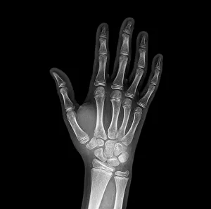
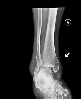
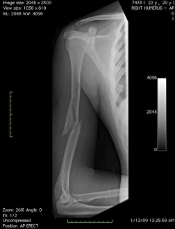
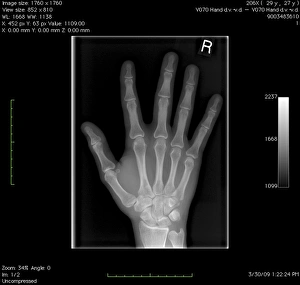
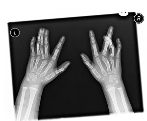
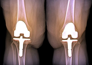
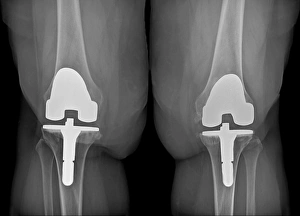
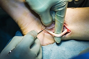
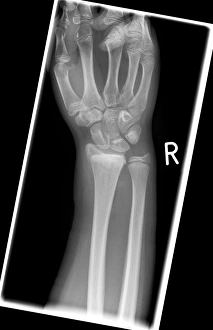
"Exploring the World of Orthopaedics: From Broken Bones to Artistic Marvels" A glimpse into orthopaedic challenges: X-ray reveals a broken wrist bone


All Professionally Made to Order for Quick Shipping
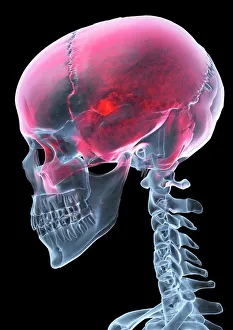
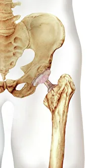
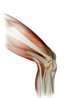
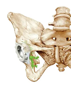
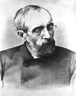
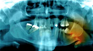
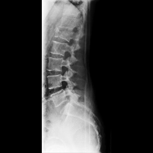
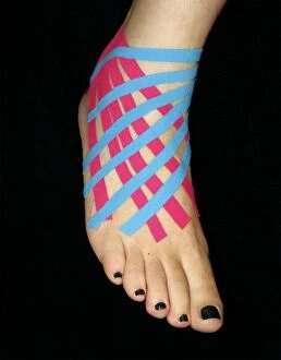
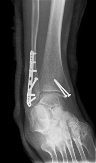
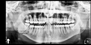
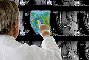
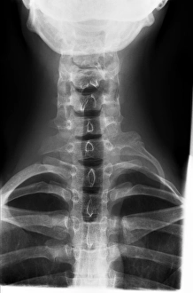
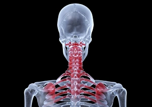
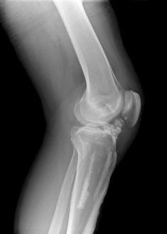
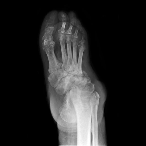
"Exploring the World of Orthopaedics: From Broken Bones to Artistic Marvels" A glimpse into orthopaedic challenges: X-ray reveals a broken wrist bone, reminding us of the intricate nature of healing. (X-ray C017 / 7187) The intersection of art and medicine: An exquisite artwork depicts the beauty of a hip replacement procedure, showcasing human resilience and innovation. Unveiling the hidden damage: Through an artistic representation, we witness the complexity of a damaged knee ligament and appreciate the expertise required for its restoration. Beyond physical pain: An intriguing X-ray artwork captures a headache, reminding us that orthopaedics encompasses not only bones but also various conditions affecting our well-being. Celebrating transformative surgeries: Another captivating artwork showcases a hip replacement, symbolizing hope and renewed mobility for countless individuals worldwide. Remembering Hugh Owen Thomas's legacy: Welsh surgeon C016 / 6299 revolutionized orthopaedics with his pioneering techniques, leaving an indelible mark on medical history. Peering beneath the surface: A jaw-dropping X-ray exposes a fractured jawbone, emphasizing both the fragility and strength inherent in our skeletal system. Historical insights into orthopaedic practices: An engraving from Traite des bandages et appareils illustrates a corset designed to prevent onanism—a testament to evolving medical beliefs in Paris during 1815 (b/w photo). Nurturing young lives with care: Dorothea Lange's poignant photograph captures a baby wearing homemade splints for club feet at an FSA camp in Tulare County, California in 1939—an early example of personalized orthopaedic treatment. Embracing knowledge through literature: The title page from L'Orthopedie ou l'Art de Prevenir et de Corriger dans les invites us to explore this comprehensive guide to preventing and correcting orthopaedic conditions.