
Proximal Collection
"Exploring the Proximal: From Kidney Tubules to Mammalian Bones and Medical Interventions" In this captivating journey through various scientific realms


All Professionally Made to Order for Quick Shipping
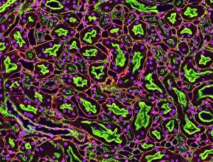
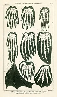
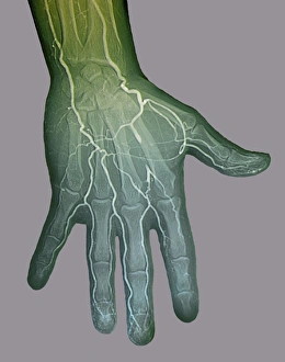
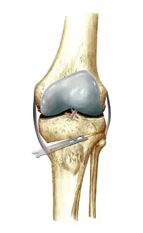
"Exploring the Proximal: From Kidney Tubules to Mammalian Bones and Medical Interventions" In this captivating journey through various scientific realms, we delve into the concept of "proximal. " Starting with a microscopic view, we observe kidney tubules in section, marveling at their intricate structure and vital role in our body's filtration system. Shifting gears, we embark on a comparative study of mammalian hands from nine different species. A stunning 1898 color lithograph showcases the diversity and complexity of these bones, highlighting nature's remarkable adaptations. Our exploration then takes us into the realm of medical imaging. Through an ischaemia digital angiogram, we witness firsthand the impact of restricted blood flow on tissues. The Paprosky femur defect classification C016/6621 sheds light on orthopedic challenges and treatment options for patients facing bone loss or damage. Next, we venture into cardiology as X-rays reveal coronary stenosis before and after treatment. These images showcase both the severity of arterial blockages and the transformative power of medical interventions in restoring blood flow to compromised hearts. Returning to orthopedics, knee osteotomy artwork C016/6995 provides a visual representation of surgical techniques used to correct misalignments or deformities in knee joints. This artistic rendering captures both technical precision and hope for improved mobility. In this captivating collection spanning diverse fields such as nephrology, mammalogy, interventional radiology, cardiology, and orthopedics – proximal emerges as a unifying theme. It reminds us that understanding structures close to us can unlock profound insights about our bodies' inner workings while offering innovative solutions for health challenges that affect millions worldwide.