
Bone Cement Collection
"Revolutionizing Orthopedic Surgeries: The Power of Bone Cement" In the ever-evolving field of orthopedics, it has emerged as a game-changer


All Professionally Made to Order for Quick Shipping
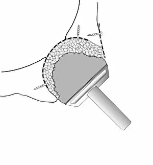
"Revolutionizing Orthopedic Surgeries: The Power of Bone Cement" In the ever-evolving field of orthopedics, it has emerged as a game-changer, providing hope and restoring mobility to countless individuals. From addressing complex femur defects to enhancing prosthetic hip joints, this remarkable substance continues to redefine the possibilities in surgical interventions. One crucial application lies in Paprosky femur defect classification (C016 / 6621). Surgeons can now effectively tackle challenging cases by utilizing this innovative solution, enabling them to reconstruct damaged femurs with precision and accuracy. By understanding the unique characteristics of each defect type, such as IIIA lateral or IIIA med-lat, medical professionals can tailor their approach for optimal outcomes. Moreover, when it comes to prosthetic hip joint surgeries (artwork C016 / 6782), bone cement plays an integral role in ensuring stability and longevity. With detailed diagrams showcasing Gruen zones (C016 / 6776) and comprehensive illustrations depicting prosthetic hip joints (diagram C016 / 6775), surgeons gain valuable insights into strategic placement techniques that enhance patient comfort and functionality. The significance of bone grafting cannot be overlooked either—particularly in hip socket procedures. Through meticulous diagrammatic representations (C016 / 6784 & C016 / 6785), we witness how bone cement facilitates successful graft integration during these intricate surgeries. This technique not only strengthens the socket but also promotes long-term implant fixation for improved patient satisfaction. Even failed hip replacements find redemption through composite images highlighting corrective measures (Failed hip replacement composite image). Thanks to advancements in bone cement technology, revision surgeries have become more effective than ever before. Surgeons can rectify previous complications with confidence while ensuring enhanced stability and durability for patients seeking a second chance at pain-free living. As we delve deeper into the world of orthopedic marvels like bone cement, one thing becomes abundantly clear.