
Hip Replacement Collection
"Exploring the Art and Science of Hip Replacement: A Journey through Time" Step into the world as we delve into its artistic representations and medical advancements


All Professionally Made to Order for Quick Shipping
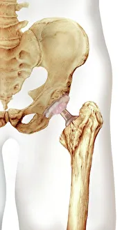
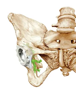
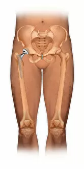
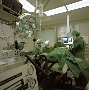
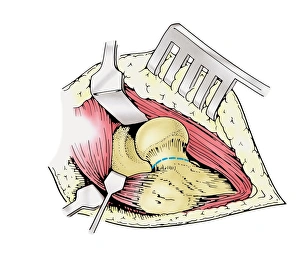
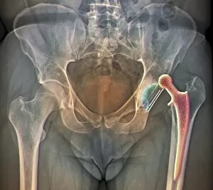
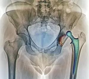
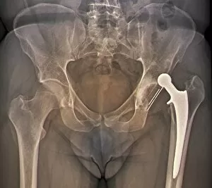
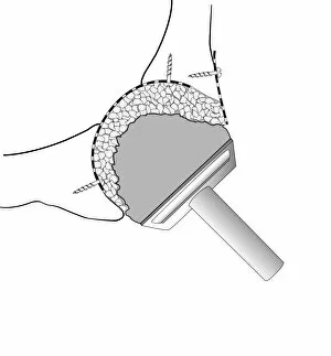
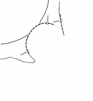
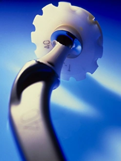
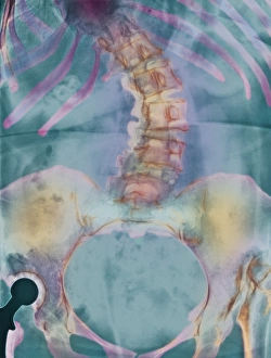
"Exploring the Art and Science of Hip Replacement: A Journey through Time" Step into the world as we delve into its artistic representations and medical advancements. 🎨🏥 Featuring captivating artwork showcasing a front view of a body with a total hip replacement, we are transported to the Royal Masonic Hospital in Hammersmith, London, circa 1980. The artist's meticulous attention to detail brings this hospital operating theatre to life, allowing us to witness the intricate procedure that revolutionized lives. As we examine Paprosky femur defect classification C016 / 6621 diagram, we gain insight into the complexities surgeons face when dealing with various bone conditions. This visual representation highlights their expertise in removing affected heads of femur bones – an essential step towards restoring mobility and alleviating pain. Zooming in on a close-up shot of a hip joint prosthesis reveals the incredible craftsmanship behind these artificial joints. Meticulously designed and engineered for durability and functionality, they seamlessly integrate with our bodies, offering renewed hope for those suffering from debilitating hip conditions. However, complications can arise even after surgery. X-ray F006 / 9132 showcases a dislocated hip replacement – reminding us that challenges may still persist along this journey towards recovery. Yet medical professionals continue to strive for excellence as evidenced by X-rays F006 / 9131 & F006 / 9130 which demonstrate their tireless efforts in addressing such issues. Finally, exploring Gruen zones C016 / 6778 allows us to understand how prosthetic hip joints interact within our bodies' unique anatomical structures. These zones serve as guidelines for surgeons during implantation procedures ensuring optimal placement and stability – crucial factors contributing to successful outcomes. Join us on this captivating exploration where art meets science; unveiling both triumphs and challenges encountered throughout the fascinating realm of hip replacements.