Fraud Blocker Skip to main content

Prostheses Collection

"Revolutionizing Lives: The Evolution of Prostheses" In the realm of history

Background imageProstheses Collection: A new way to pay the National Debt, pub. 21st April, 1786 (hand coloured engraving)

A new way to pay the National Debt, pub. 21st April, 1786 (hand coloured engraving)
2636952 A new way to pay the National Debt, pub. 21st April, 1786 (hand coloured engraving) by Gillray, James (1757-1815); Private Collection; (add.info)

Background imageProstheses Collection: Paprosky femur defect classification C016 / 6621

Paprosky femur defect classification C016 / 6621
Paprosky femur defect classification. Diagram showing the classification system for femur cortex defects used to assess revision (replacement or repair) of a hip implant

Background imageProstheses Collection: A new way to pay the national-debt, Cartoon shows King George III and Queen Charlotte

A new way to pay the national-debt, Cartoon shows King George III and Queen Charlotte standing before the Treasury, moneybags under their arms

Background imageProstheses Collection: A New Way To Pay The National-debt

A New Way To Pay The National-debt, Cartoon Shows King George Iii And Queen Charlotte Standing Before The Treasury, Moneybags Under Their Arms

Background imageProstheses Collection: Total knee replacement, X-ray

Total knee replacement, X-ray
Total knee replacement. X-ray of the legs of a 49-year-old patient who has had a total knee replacement (white) of the left knee (right)

Background imageProstheses Collection: Prosthetic hip joint and Gruen zones C016 / 6778

Prosthetic hip joint and Gruen zones C016 / 6778
Prosthetic hip joint and Gruen zones. Cutaway diagram of a femur (thigh bone) showing a femoral component of a hip prosthesis and labels indicating Gruen zones

Background imageProstheses Collection: Prosthetic hip joints and Gruen zones C016 / 6781

Prosthetic hip joints and Gruen zones C016 / 6781
Prosthetic hip joints and Gruen zones. Diagram of the femoral components of two types of hip prosthesis with labels indicating standard and modified Gruen zones

Background imageProstheses Collection: Prosthetic hip joint, artwork C016 / 6782

Prosthetic hip joint, artwork C016 / 6782
Prosthetic hip joint. Cutaway artwork of a femur (thigh bone) showing a femoral component of a hip prosthesis. This component is implanted in the femur after the head of the femur has been surgically

Background imageProstheses Collection: Artificial heart valves, artwork C016 / 7495

Artificial heart valves, artwork C016 / 7495
Artificial heart valves, computer artwork. There are four valves in the human heart, all of which prevent the backflow of blood

Background imageProstheses Collection: Hip socket bone grafting, diagram C016 / 6786

Hip socket bone grafting, diagram C016 / 6786
Hip socket bone grafting. Diagram showing completed hip socket bone grafting as part of a total hip replacement operation

Background imageProstheses Collection: Hip socket bone grafting, diagram C016 / 6784

Hip socket bone grafting, diagram C016 / 6784
Hip socket bone grafting. Diagram showing the second stage in hip socket bone grafting as part of a total hip replacement operation

Background imageProstheses Collection: Hip socket bone grafting, diagram C016 / 6785

Hip socket bone grafting, diagram C016 / 6785
Hip socket bone grafting. Diagram showing the third stage in hip socket bone grafting as part of a total hip replacement operation

Background imageProstheses Collection: Hip socket bone grafting, diagram C016 / 6783

Hip socket bone grafting, diagram C016 / 6783
Hip socket bone grafting. Diagram showing the first stage in hip socket bone grafting as part of a total hip replacement operation

Background imageProstheses Collection: Prosthetic hip joint, diagram C016 / 6775

Prosthetic hip joint, diagram C016 / 6775
Prosthetic hip joint. Cutaway diagram of a femur (thigh bone) showing a femoral component of a hip prosthesis. This component is implanted in the femur after the head of the femur has been surgically

Background imageProstheses Collection: Prosthetic hip joint and Gruen zones C016 / 6777

Prosthetic hip joint and Gruen zones C016 / 6777
Prosthetic hip joint and Gruen zones. Cutaway diagram of a femur (thigh bone) showing a femoral component of a hip prosthesis and labels indicating Gruen zones

Background imageProstheses Collection: Prosthetic hip joint and Gruen zones C016 / 6780

Prosthetic hip joint and Gruen zones C016 / 6780
Prosthetic hip joint and Gruen zones. Diagram of the femoral component of a hip prosthesis with labels indicating the hip connection (upper left) and the seven Gruen zones

Background imageProstheses Collection: Prosthetic hip joint and Gruen zones C016 / 6776

Prosthetic hip joint and Gruen zones C016 / 6776
Prosthetic hip joint and Gruen zones. Cutaway diagram of a femur (thigh bone) showing a femoral component of a hip prosthesis and labels indicating Gruen zones

Background imageProstheses Collection: Prosthetic hip joint and Gruen zones C016 / 6779

Prosthetic hip joint and Gruen zones C016 / 6779
Prosthetic hip joint and Gruen zones. Cutaway diagram of a femur (thigh bone) showing a femoral component of a hip prosthesis and labels indicating Gruen zones

Background imageProstheses Collection: Double mastectomy breast implants, MRI

Double mastectomy breast implants, MRI
Double mastectomy breast implants. Post-operative coloured magnetic resonance imaging (MRI) scan of a section through the chest of a 61-year-old patient after a double mastectomy (breast removal)

Background imageProstheses Collection: Early 19th Century facial prosthetics

Early 19th Century facial prosthetics. Selection of false eyes, ears, noses and spectacles from 1920

Background imageProstheses Collection: Breast implants

Breast implants. Silicone implants are used in several types of cosmetic surgery, most commonly for surgery to enlarge a breast or to restore breast shape after breast tissue removal for cancer

Background imageProstheses Collection: Bionic limbs, artwork

Bionic limbs, artwork
Bionic limbs. Computer artwork or a man whose right arm and leg have been replaced with robotic prostheses that are controlled by his brain

Background imageProstheses Collection: Artificial knee joints in obesity, X-ray

Artificial knee joints in obesity, X-ray
Artificial knee joints in obesity. X-ray of the replacement knee joints of an obese patient. This is a weight-bearing view from the front

Background imageProstheses Collection: Paprosky femur defect, type IIIA lateral

Paprosky femur defect, type IIIA lateral
Paprosky femur defect. Cutaway artwork of bone degradation in a type IV medial-lateral femur cortex defect (Paprosky classification system)

Background imageProstheses Collection: Paprosky femur defect, type IIIA med-lat

Paprosky femur defect, type IIIA med-lat
Paprosky femur defect. Cutaway artwork of bone degradation in a type IV lateral femur cortex defect (Paprosky classification system)

Background imageProstheses Collection: Knee replacement surgery C017 / 8547

Knee replacement surgery C017 / 8547
Knee replacement surgery. Prosthetic knee implants ready for use in total knee replacement surgery. During the operation prostheses will be implanted into the thigh bone (femur) and shinbone (tibia)

Background imageProstheses Collection: Medical technology

Medical technology

Background imageProstheses Collection: Technological hand

Technological hand. Conceptual image of a hand and technological equipment. This could represent the development of technology being in part due to the dexterity of a human hand

Background imageProstheses Collection: Cyborg running

Cyborg running, computer artwork. A cyborg or bionic man is an entity that is part man and part machine

Background imageProstheses Collection: Double hip replacement, X-ray

Double hip replacement, X-ray
Double hip replacement. Coloured X-ray of a female patients hips with two prosthetic hip implants (white). Each implant is embedded in a hole drilled into its femur (thigh bone)

Background imageProstheses Collection: Heart pacemaker, X-ray

Heart pacemaker, X-ray
Heart pacemaker. Coloured chest X-ray showing a surgically-implanted heart pacemaker (red, centre right). The pacemaker supplies electrical impulses along leads (running to lower centre)

Background imageProstheses Collection: Selection of 16th century artificial arms & hands

Selection of 16th century artificial arms & hands
Engraving of a selection of artificial hands & arms devised & drawn by the French surgeon Ambroise Pare (1510-1590). Pare attached himself to the army as a barber-surgeon

Background imageProstheses Collection: Breast implant calcification, X-ray

Breast implant calcification, X-ray
Breast implant calcification. Coloured mammogram (breast X-ray) of calcification around the borders of a breast implant (pink) in a womans breast (red) as seen from the side

Background imageProstheses Collection: Fusion of spinal bones, X-ray

Fusion of spinal bones, X-ray
Coloured X-ray (front view) of the spine in a 45 year old patient with degenerative disc disease showing two implants (prostheses) between vertebral bones L4-L5 and L5-S1

Background imageProstheses Collection: Prosthetic shafts for hip replacement

Prosthetic shafts for hip replacement
Prosthetic shaft for hip replacement. These prostheses are inserted into the femur (thigh bone). They will have a ball attached at the top, which will form part of the hip joint


All Professionally Made to Order for Quick Shipping

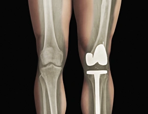
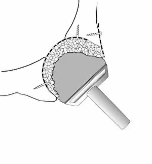
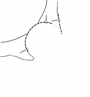
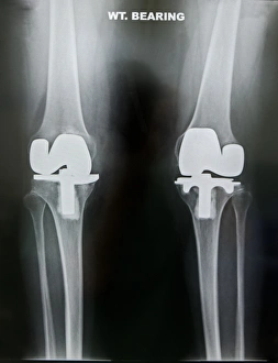
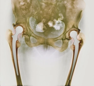
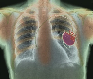
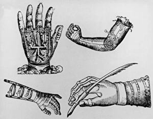
"Revolutionizing Lives: The Evolution of Prostheses" In the realm of history, a hand-coloured engraving titled "A new way to pay the National Debt" was published on April 21st, 1786. Little did they know that this artwork would foreshadow an incredible journey into the world of prostheses. Fast forward to modern times, where medical advancements have brought forth remarkable solutions for individuals facing physical challenges. One such innovation is the Paprosky femur defect classification C016/6621 – a groundbreaking technique that has revolutionized total knee replacements. With its precision and effectiveness, it offers hope and improved mobility to countless patients. Similarly, we delve into a satirical cartoon named "A new way to pay the national-debt, " depicting King George III and Queen Charlotte. While amusing in nature, this artwork subtly hints at how prosthetic technology can alleviate financial burdens by enabling individuals with disabilities to contribute actively to society. The concept of paying debts takes another turn as we explore artificial heart valves depicted in captivating artwork C016/7495. These life-saving devices showcase how science merges with artistry to create miracles within our bodies – mending hearts and restoring vitality. Delving deeper into orthopedic wonders, we encounter diagrams illustrating hip socket bone grafting (C016/6786 & C016/6784). These intricate procedures highlight how medical professionals utilize cutting-edge techniques to restore functionality and enhance quality of life for those suffering from hip joint issues. Lastly, let us not forget about prosthetic hip joints (C016/6778 & C016/6781) which have become synonymous with freedom for many individuals worldwide. Through these marvels of engineering combined with Gruen zones analysis techniques, countless lives are transformed as people regain their independence and rediscover joy in everyday activities once again.