
Arthrology Collection
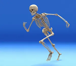
"Exploring the Intricate World of Arthrology: Unveiling the Secrets Behind Our Body's Skeleton" Running skeleton in body


All Professionally Made to Order for Quick Shipping
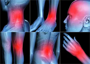
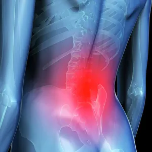
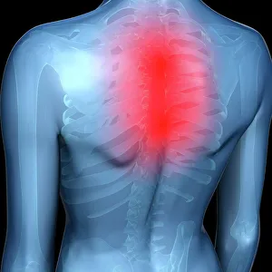
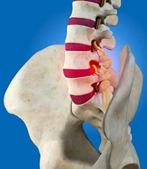
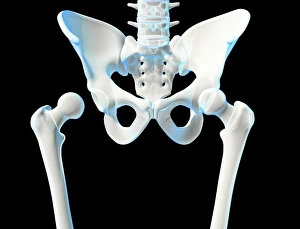
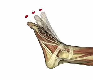
"Exploring the Intricate World of Arthrology: Unveiling the Secrets Behind Our Body's Skeleton" Running skeleton in body, artwork: Witness the graceful motion of a running skeleton, as arthrology unravels the hidden beauty within our bodies. Body pain, artwork: Dive into an artistic representation of body pain, where arthrology sheds light on understanding and alleviating discomfort. Lower back pain, conceptual artwork: Delve into a conceptual masterpiece that depicts lower back pain and how arthrology plays a crucial role in diagnosing and treating this common ailment. Upper back pain, conceptual artwork: Explore an intriguing concept art piece illustrating upper back pain and how arthrology helps us comprehend its underlying causes. Outer ankle ligaments, artwork C013 / 4452: Discover the intricacies of outer ankle ligaments through captivating artwork that showcases their importance in maintaining stability during movement. Inner ankle ligaments, artwork C013 / 4451: Journey deep inside our ankles with mesmerizing artistry that highlights inner ankle ligaments' significance in supporting our every step. Slipped intervertebral disc, artwork: Get lost in a stunning visual representation of a slipped intervertebral disc—a condition understood by arthrologists who strive to restore comfort and mobility to affected individuals. Male skeleton: Marvel at the elegance of a male skeleton captured artistically while contemplating how arthrology unveils insights about bone structure and joint function across genders. Neck pain, conceptual artwork: Immerse yourself in thought-provoking imagery portraying neck pain—an affliction often studied by experts well-versed in arthrological knowledge for effective treatment strategies. Hip joint bones and anatomy, artwork C014 / 2032: Embark on an educational journey exploring hip joint bones' intricate anatomy through visually striking illustrations crafted by arthrology enthusiasts.