Fine Art Print > Fine Art Storehouse > Digital Vision Vectors > Science
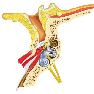
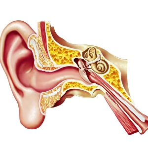
Fine Art Print : Anatomy of the human ear, lithograph, published in 1876


![]()
Fine Art Prints from Fine Art Storehouse
Anatomy of the human ear, lithograph, published in 1876
Anatomy of the human ear. Lithograph, published in 1876
Unleash your creativity and transform your space into a visual masterpiece!
ZU_09
Media ID 18292415
© ZU_09
Cochlea Ear Drum Incus Malleus Membrane Middle Ear Stapes
20"x16" (+3" Border) Fine Art Print
Discover the intricacies of the human body with this stunning lithograph, Anatomy of the Human Ear, from the Fine Art Storehouse collection. Published in 1876 by ZU_09, this fine art print showcases the complex structure of the ear in exquisite detail. A must-have for anatomy enthusiasts, medical professionals, and art collectors alike, this historical piece adds depth and character to any space. Bring the beauty of science into your home or office with this captivating and educational print.
20x16 image printed on 26x22 Fine Art Rag Paper with 3" (76mm) white border. Our Fine Art Prints are printed on 300gsm 100% acid free, PH neutral paper with archival properties. This printing method is used by museums and art collections to exhibit photographs and art reproductions.
Our fine art prints are high-quality prints made using a paper called Photo Rag. This 100% cotton rag fibre paper is known for its exceptional image sharpness, rich colors, and high level of detail, making it a popular choice for professional photographers and artists. Photo rag paper is our clear recommendation for a fine art paper print. If you can afford to spend more on a higher quality paper, then Photo Rag is our clear recommendation for a fine art paper print.
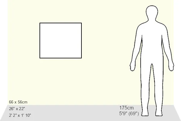
Estimated Image Size (if not cropped) is 50.8cm x 40.6cm (20" x 16")
Estimated Product Size is 66cm x 55.9cm (26" x 22")
These are individually made so all sizes are approximate
Artwork printed orientated as per the preview above, with landscape (horizontal) orientation to match the source image.
EDITORS COMMENTS
This lithograph, published in 1876, offers a detailed glimpse into the intricate anatomy of the human ear. Created by the talented artist ZU_09, this print showcases their remarkable skill in capturing scientific subjects with artistic finesse. The composition is meticulously crafted, presenting an array of anatomical structures that make up this vital sensory organ. At the center of attention lies the cochlea, resembling a delicate seashell spiraling gracefully within the frame. Surrounding it are various components such as the stapes and malleus bones, which form part of the middle ear mechanism responsible for transmitting sound vibrations to our brain. The ear drum, or tympanic membrane, takes prominence as a translucent barrier separating external sounds from our inner auditory system. ZU_09's lithograph beautifully combines scientific accuracy with aesthetic appeal. The fine lines and meticulous shading bring each element to life while maintaining an air of elegance throughout. This artwork serves not only as a visual representation but also as an educational tool that allows us to explore and appreciate the wonders hidden within our own bodies. Whether displayed in a medical office or art enthusiast's collection, this print undoubtedly sparks curiosity and admiration for both science and art lovers alike. It stands as a testament to ZU_09's talent in merging technical precision with artistic expression—a timeless piece that continues to captivate viewers over a century after its creation.
MADE IN THE USA
Safe Shipping with 30 Day Money Back Guarantee
FREE PERSONALISATION*
We are proud to offer a range of customisation features including Personalised Captions, Color Filters and Picture Zoom Tools
SECURE PAYMENTS
We happily accept a wide range of payment options so you can pay for the things you need in the way that is most convenient for you
* Options may vary by product and licensing agreement. Zoomed Pictures can be adjusted in the Cart.