
Stapes Collection
"Unveiling the Intricacies of the Human Ear: A Journey through Stapes" Step back in time to 1876 as we explore the fascinating anatomy of the human ear


All Professionally Made to Order for Quick Shipping
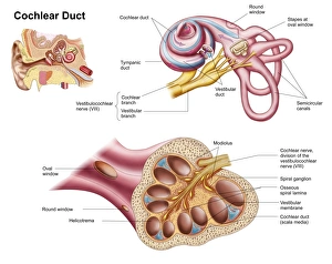
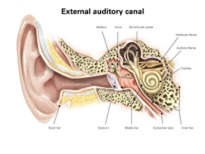
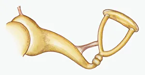
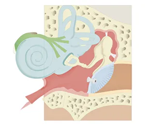
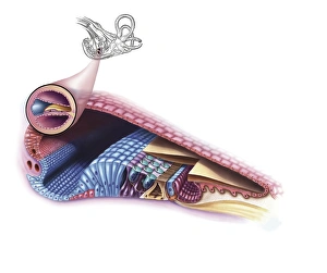
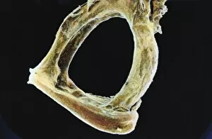
"Unveiling the Intricacies of the Human Ear: A Journey through Stapes" Step back in time to 1876 as we explore the fascinating anatomy of the human ear. This lithograph, published over a century ago, takes us on a visual tour of this intricate sensory organ. Delving deeper into our exploration, we encounter the cochlear duct within the human ear. Its complex structure is unveiled before our eyes, showcasing its vital role in hearing and balance. Moving towards the external auditory canal, labeled with precision, we gain insight into how sound waves enter this remarkable system. The significance of this pathway becomes apparent as we unravel more about our auditory capabilities. The cutaway diagram presents an awe-inspiring view of the entire human ear. Every component meticulously illustrated to showcase its function and interconnection within this marvel of nature. Zooming closer into focus, we witness an exquisite illustration capturing one particular bone - stapes. Resembling a stirrup shape within the middle ear of Epitheria mammals, it plays a crucial role in transmitting sound vibrations from eardrum to inner ear structures. A digital masterpiece reveals not only stapes but also its companions - malleus and incus - forming a chain that amplifies sound signals for optimal perception. This intricate arrangement showcases nature's ingenuity at work. As we venture further inward, another digital illustration unravels both middle and inner ear complexities. Surrounded by delicate membranes and fluid-filled chambers lies an extraordinary world responsible for transforming vibrations into meaningful sounds. Taking yet another close-up look at these incredible bones – malleus, incus, and stapes – their detailed depiction captivates us once again. These tiny ossicles demonstrate their pivotal role in conducting sound waves towards our brain for interpretation. Cross-section biomedical illustrations provide invaluable insights into various aspects of our ears' functionality.