Home > Popular Themes > Human Body
Spondylodiscitis, MRI scan
![]()

Wall Art and Photo Gifts from Science Photo Library
Spondylodiscitis, MRI scan
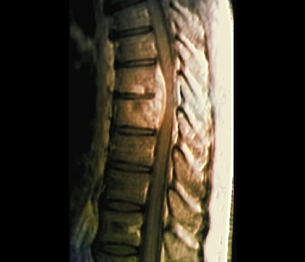
Spondylodiscitis. Coloured magnetic resonance imaging (MRI) scan of a sagittal section through the spine of a 69-year-old patient with spondylodiscitis at the level of the T7-T8 vertebra, causing compression of the spinal cord (upper centre). Spondylodiscitis is a combination of discitis (inflammation of one or more intervertebral disc spaces) and spondylitis (inflammation of one or more vertebrae)
Science Photo Library features Science and Medical images including photos and illustrations
Media ID 9247435
© ZEPHYR/SCIENCE PHOTO LIBRARY
Backbone Central Nervous System Compressed Compression Diagnostic Imaging Discs Inflamed Inflammation Intervertebral Disc Magnetic Resonance Imaging Orthopaedic Orthopaedics Orthopedic Orthopedics Radiography Radiological Radiology Sagittal Scan Sections Seventies Spaces Spinal Spinal Cord Thoracic Vertebra Vertebral Abnormal Condition Disorder Neurological Neurology Section Sectioned Unhealthy Vertebrae
EDITORS COMMENTS
This print from Science Photo Library showcases a coloured magnetic resonance imaging (MRI) scan of a sagittal section through the spine of a 69-year-old patient with spondylodiscitis. The condition is observed at the level of the T7-T8 vertebra, resulting in compression of the spinal cord visible at the upper center of the image. Spondylodiscitis is an amalgamation of discitis, which refers to inflammation within intervertebral disc spaces, and spondylitis, characterized by inflammation affecting one or more vertebrae. The black background intensifies our focus on this medical anomaly that affects both biology and medicine. This detailed anatomical representation reveals an unhealthy state where the vertebrae are compressed and inflamed, causing disorder within the spine. The radiography highlights abnormalities in this specific region of the human body's central nervous system (CNS). Through advanced diagnostic imaging techniques like MRI scans, healthcare professionals can accurately assess conditions such as spondylodiscitis. In orthopedics and neurology alike, understanding these intricate sections allows for effective treatment plans tailored to each patient's needs. This photograph serves as a reminder of how medical advancements enable us to delve deep into our biological structures and uncover ailments that were once hidden mysteries. It emphasizes both scientific progress and compassionate care for individuals experiencing complex spinal disorders like spondylodiscitis.
MADE IN THE USA
Safe Shipping with 30 Day Money Back Guarantee
FREE PERSONALISATION*
We are proud to offer a range of customisation features including Personalised Captions, Color Filters and Picture Zoom Tools
SECURE PAYMENTS
We happily accept a wide range of payment options so you can pay for the things you need in the way that is most convenient for you
* Options may vary by product and licensing agreement. Zoomed Pictures can be adjusted in the Cart.


