
Diagnostic Imaging Collection
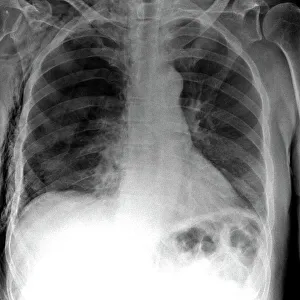
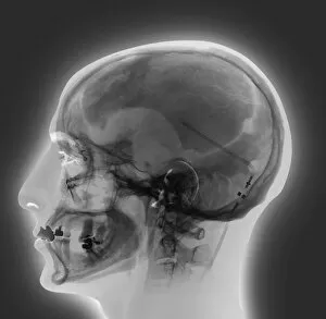
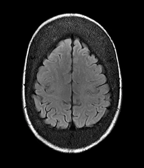
"Unlocking the Secrets of the Body: Exploring Diagnostic Imaging" Revealing Hidden Dangers: Tension pneumothorax detected through an X-ray


All Professionally Made to Order for Quick Shipping
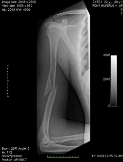
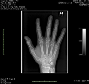
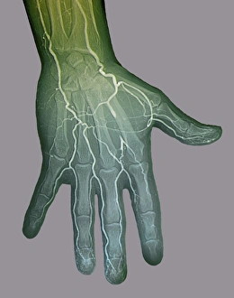
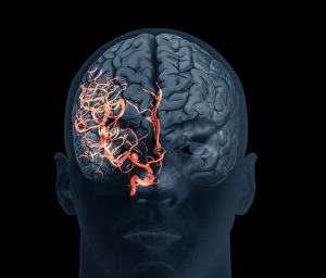
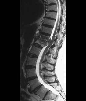
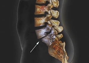
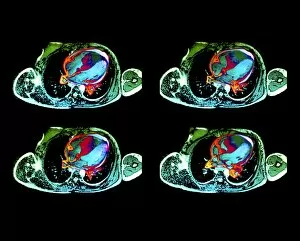
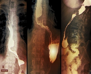
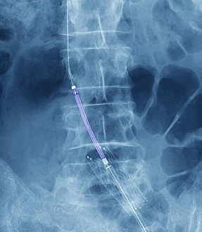
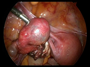
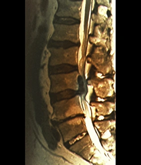
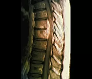
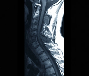
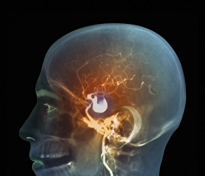
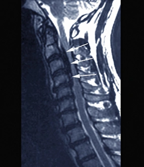
"Unlocking the Secrets of the Body: Exploring Diagnostic Imaging" Revealing Hidden Dangers: Tension pneumothorax detected through an X-ray, ensuring prompt treatment and saving lives. Peering into the Depths: Bacterial meningitis captured by an MRI scan, aiding in accurate diagnosis and timely intervention. Unveiling Ovarian Mysteries: Endoscope view C017 / 6800 exposes an ovarian cyst, guiding surgeons towards effective treatment options. Journey Inside the Uterus: Endoscope view C017 / 6805 offers a unique perspective on uterine health, enabling early detection of abnormalities. Visualizing Fractures with Precision: Digital X-ray captures a broken arm bone from multiple angles, assisting orthopedic specialists in devising appropriate treatment plans. A Glimpse of Normalcy: Digital X-ray showcases a healthy hand structure for comparative analysis and reference purposes. Mapping Blood Flow Anomalies: Ischaemia visualized through digital angiogram aids vascular experts in identifying blockages and planning interventions accordingly. Navigating Complex Brain Terrain: Cutting-edge 3D scan reveals brain aneurysm details crucial for neurosurgeons to plan precise surgical procedures or other treatments. Illuminating Spinal Infections: Tuberculosis of the spine exposed via MRI scan helps physicians initiate targeted therapies for better patient outcomes. Unmasking Spinal Misalignment Woes: Spondylolisthesis unraveled through a comprehensive 3D CT scan assists orthopedic specialists in designing personalized treatment strategies for patients' relief and recovery. Decoding Cardiac Challenges with Clarity: MRI scans unravel cardiac lymphoma's intricate manifestations within heart tissues, facilitating tailored therapeutic approaches to combat this rare condition effectively. Capturing Silent Threats Within Throat Walls:X-rays uncover hidden signs of throat cancer, enabling early detection and potentially life-saving interventions.