
Inflamed Collection
"Inflamed: A Visual Journey through the Depths of Pain" Step into a world where pain takes on an artistic form, revealing the hidden struggles within our bodies


All Professionally Made to Order for Quick Shipping
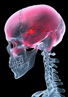
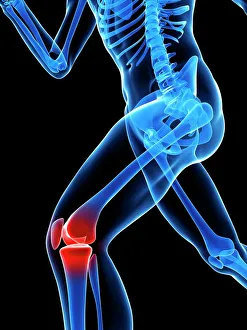
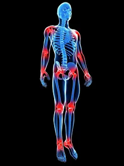
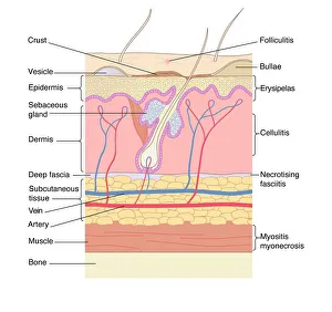
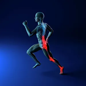
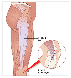
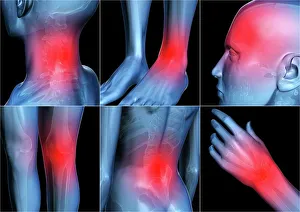
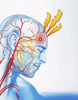
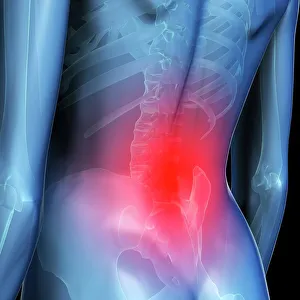
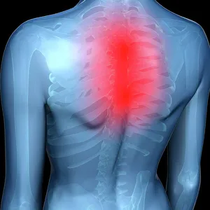
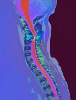
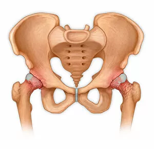
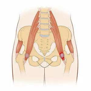
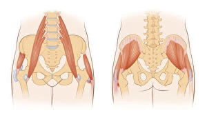
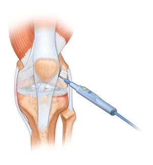
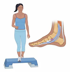
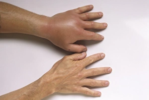
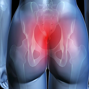
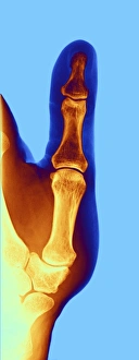
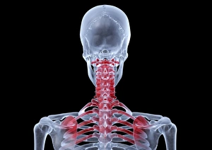
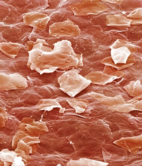
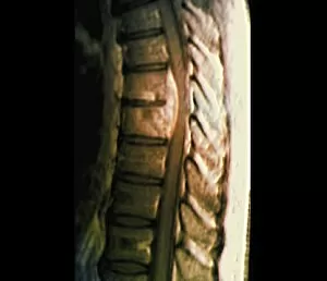
"Inflamed: A Visual Journey through the Depths of Pain" Step into a world where pain takes on an artistic form, revealing the hidden struggles within our bodies. From knee pain to joint discomfort, each artwork captures the essence of inflammation in its own unique way. In one piece, a conceptual artwork portrays knee pain as fiery red strokes intertwining with delicate brushstrokes, symbolizing the intensity and persistence of this common ailment. Similarly, another artwork depicts joint pain as a chaotic blend of colors and shapes that mirror the tumultuous nature of this condition. Moving beyond physical discomfort, skin disorders come alive through intricate artwork. The canvas becomes a battleground for vibrant hues and textured layers that reflect the agony experienced by those grappling with these conditions. A mesmerizing X-ray artwork delves into the realm of headaches, showcasing intricate patterns radiating from within. This visual representation serves as a reminder that even unseen ailments can have profound effects on our well-being. Running injuries take center stage in conceptual artworks that capture both their physical toll and emotional weight. Each stroke conveys resilience amidst adversity while highlighting the importance of self-care in preventing such setbacks. The specific agony caused by iliotibial running injury finds expression in an evocative piece where sharp lines intersect against a backdrop drenched in shades of blue – capturing both movement and restriction simultaneously. Body pain is given life through abstract artistry; bold splashes convey anguish while subtle details hint at hope for relief. Meanwhile, lower back pain emerges as ethereal swirls dancing across canvases - representing both fragility and strength intertwined. Upper back pain takes shape through conceptual pieces that juxtapose sharp angles with soft curves - mirroring how this affliction disrupts daily life yet also offers opportunities for growth and healing. Migraine sufferers find solace within vivid paintings where bursts of color mimic throbbing sensations behind closed eyes. These captivating visuals offer empathy to those who battle these debilitating headaches.