
Thoracic Collection
"Exploring the Marvels of Thoracic: Unveiling the Secrets of the Heart and Beyond" Delve into the intricate world anatomy


All Professionally Made to Order for Quick Shipping
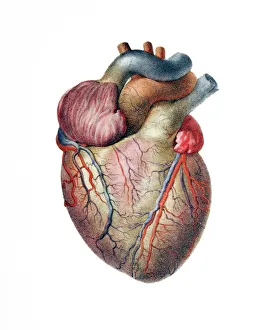
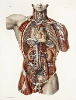
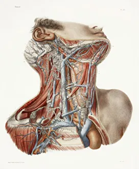
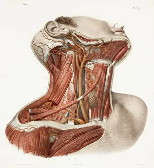
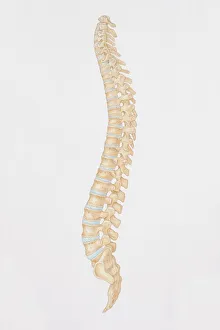
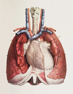
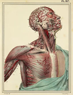
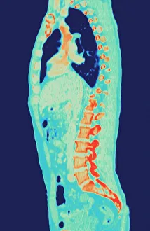
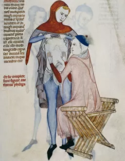
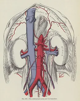
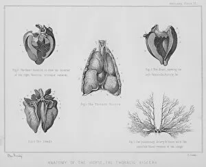
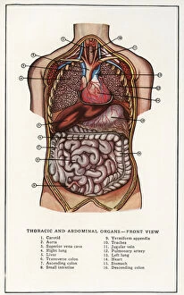
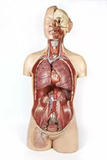
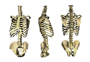
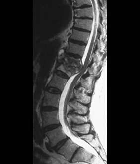
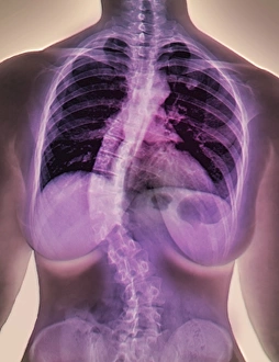
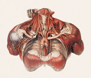
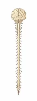
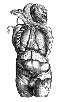
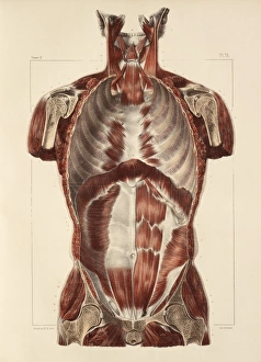
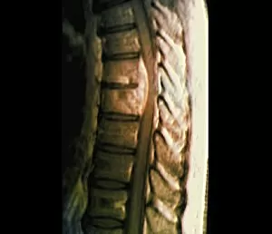
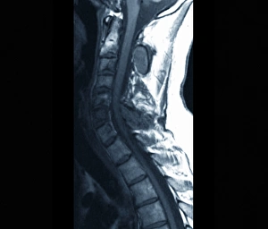
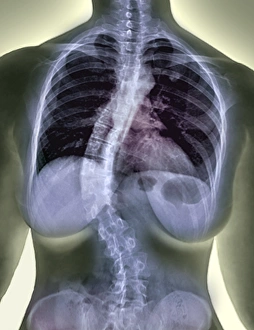
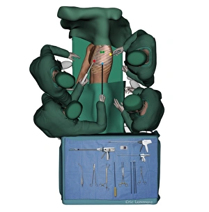
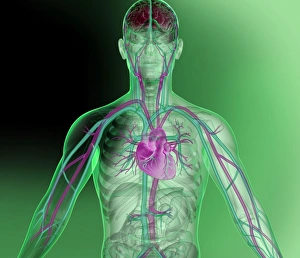
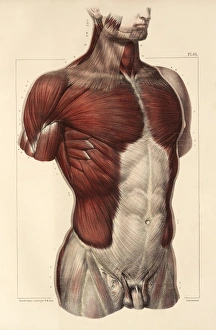
"Exploring the Marvels of Thoracic: Unveiling the Secrets of the Heart and Beyond" Delve into the intricate world anatomy, where the heart reigns as its majestic centerpiece. Journey through centuries past with historical artwork showcasing the cardiovascular system's captivating beauty. Admire 19th-century masterpieces depicting neck anatomy, offering a glimpse into our complex vascular network. Behold mesmerizing historical artwork unraveling the intricacies of neck vascular anatomy like never before seen. Discover a side view diagram unveiling the human spine's remarkable structure within the thoracic region. Witness how heart and lungs harmoniously coexist in this vital part of our body, ensuring life's rhythm continues to beat. Step back in time with enchanting 1825 artwork illustrating head and chest arteries, revealing their crucial role in sustaining life. Peer inside an MRI scan capturing a normal torso, providing invaluable insights into thoracic health assessment techniques. Marvel at another stunning MRI scan showcasing a detailed image of a human torso, unravelling its hidden mysteries layer by layer. Observe a dedicated doctor conducting thorough thoracic and abdominal recognition on their patient for comprehensive care delivery. Explore an engraving that showcases the intricate branches stemming from the abdominal aorta - nature's masterpiece at work. Immerse yourself in lithographic artistry portraying horse anatomy; uncovering secrets about thoracic viscera shared between species. Embark on this captivating journey through history and modern medicine as we unlock fascinating revelations about "Thoracic, " taking you deep into realms unseen until now.