Home > Popular Themes > Human Body
Intestinal villi anatomy, artwork

![]()
Wall Art and Photo Gifts from Science Photo Library
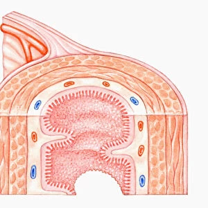
Intestinal villi anatomy, artwork
Intestinal villi anatomy. Artwork showing six types of cell found on the surface of villi in the small intestine. Clockwise from upper right they are: enterocytes (green, brush border absorptive cells, most common), enteroendocrine cells (orange), goblet cells (purple), Paneth cells (yellow), stem cells (dark beige) and proliferative progenitor cells (light beige). The latter three are at the base of the villi, while the other three are higher up, formed as stem cells mature and move up the villi. Inside the villi are blood vessels (arterial and venous capillaries, red and blue) to remove absorbed nutrients from digested food, and a lymph vessel (yellow)
Science Photo Library features Science and Medical images including photos and illustrations
Media ID 6326751
© ART FOR SCIENCE/SCIENCE PHOTO LIBRARY
Arterial Brush Border Capillaries Capillary Color Colour Colours Digestive System Enterocyte Gastroenterology Gastrointestinal Tract Gland Glandular Goblet Cell Histological Histology Intestinal Intestinal Villi Intestines Longitudinal Lymph Vessel Lymphatic System Mucosa Physiological Physiology Small Intestine Stem Cell Sub Mucosa Tissue Venous Villi Villus Cells Section Sectioned
EDITORS COMMENTS
This artwork showcases the intricate anatomy of intestinal villi found in the small intestine. The print highlights six distinct types of cells that make up the surface of these finger-like projections. Starting from the upper right and moving clockwise, we can observe enterocytes, which are green brush border absorptive cells and the most abundant type. Enteroendocrine cells appear in a vibrant orange hue, while goblet cells stand out in a regal shade of purple. Paneth cells, responsible for immune defense, shine brightly in yellow. At the base of the villi, we find stem cells (dark beige) and proliferative progenitor cells (light beige), both crucial for tissue regeneration and growth. As stem cells mature and ascend along the villi's length, they differentiate into various cell types. The artwork also provides insight into what lies within these remarkable structures. Blood vessels colored red and blue represent arterial and venous capillaries respectively; their purpose is to extract absorbed nutrients from digested food. Additionally, a lymph vessel depicted in yellow demonstrates how waste products are transported away. This visually stunning illustration not only serves as an educational tool but also emphasizes the complexity and beauty present within our own bodies. It offers a glimpse into one aspect of human biology that often goes unnoticed yet plays a vital role in our digestive system - making it truly awe-inspiring!
MADE IN THE USA
Safe Shipping with 30 Day Money Back Guarantee
FREE PERSONALISATION*
We are proud to offer a range of customisation features including Personalised Captions, Color Filters and Picture Zoom Tools
SECURE PAYMENTS
We happily accept a wide range of payment options so you can pay for the things you need in the way that is most convenient for you
* Options may vary by product and licensing agreement. Zoomed Pictures can be adjusted in the Cart.