
Capillary Collection
"Exploring the Intricate Network of Capillaries: A Journey through the Human Digestive System and Artistic Renderings" Step into the mesmerizing world of capillaries


All Professionally Made to Order for Quick Shipping
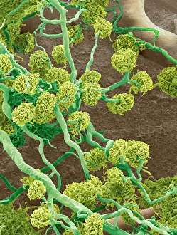
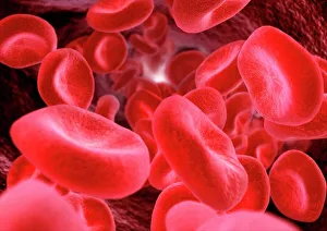
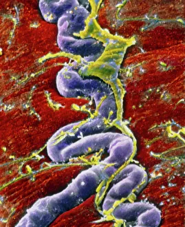
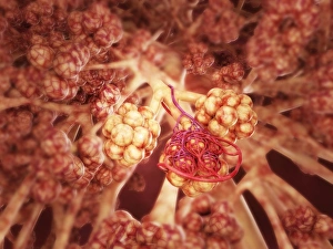
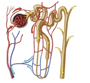
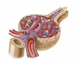
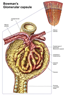
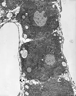
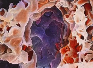
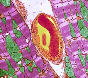
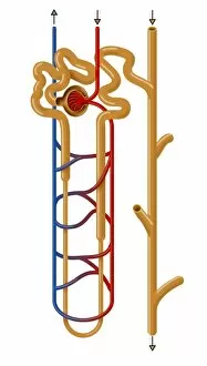
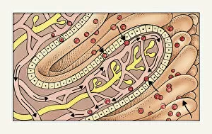
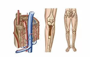
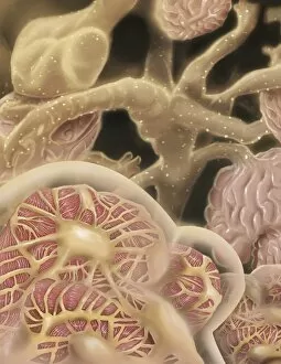
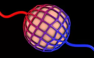
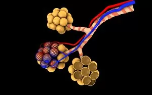
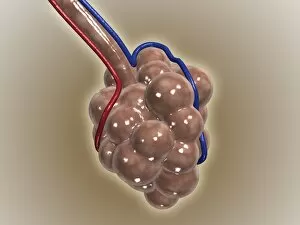
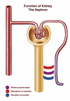
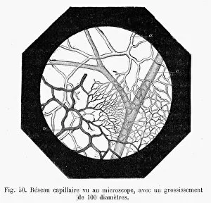
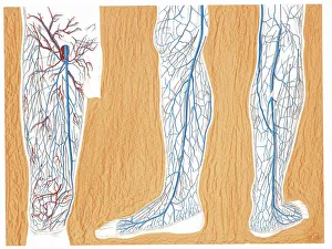
"Exploring the Intricate Network of Capillaries: A Journey through the Human Digestive System and Artistic Renderings" Step into the mesmerizing world of capillaries, where intricate pathways weave their way through our bodies. In this captivating journey, we delve into various organs and tissues to uncover the beauty and functionality of these tiny blood vessels. Starting with a closer look at the kidney glomerulus under a scanning electron microscope (SEM), we witness its complex structure that filters waste products from our blood. The delicate web-like arrangement of capillaries ensures efficient filtration, enabling our kidneys to maintain proper balance in our body. Moving on to another SEM image, we discover thyroid gland capillaries intricately intertwined within this vital endocrine organ. These microscopic vessels play a crucial role in delivering hormones throughout our body, ensuring proper metabolism and growth. As we continue exploring kidney glomeruli under SEM once again, their unique architecture comes into focus. These clusters of capillaries enable selective reabsorption and excretion processes necessary for maintaining fluid balance in our bodies. Shifting gears slightly, we turn towards retina blood vessels captured by SEM. Here, these tiny capillaries supply oxygen-rich blood to nourish the light-sensitive cells responsible for vision – an exquisite example of nature's design meeting function. Intriguingly, computer artwork showcases red blood cells flowing gracefully through capillary networks. This imaginative representation reminds us how essential these vessels are in transporting oxygen and nutrients while removing waste products throughout every corner of our bodies. Diving deeper into muscle tissue with false-color SEM imagery reveals pericyte-covered capillaries. Pericytes act as guardians protecting these fragile vessels while also playing a role in regulating blood flow – an elegant symbiotic relationship between cells within us. Concluding this captivating journey is yet another glimpse at kidney glomeruli but now observed through light microscopy. This traditional technique highlights their distinct appearance and allows us to appreciate the intricate details of these vital structures.