Fraud Blocker Skip to main content

Gland Collection

"Gland: The Hidden Marvels of Nature and Anatomy" 1) Botanik Digitalis purpurea L

Background imageGland Collection: Botanik Digitalis purpurea L. Fingerhut 160: 1

Botanik Digitalis purpurea L. Fingerhut 160: 1
Foxglove leaf. Coloured scanning electron micrograph (SEM) of a section through a leaf from a foxglove (Digitalis purpurea) plant, showing its hairs (trichomes)

Background imageGland Collection: Lactating breast tissue, light micrograph

Lactating breast tissue, light micrograph
Lactating breast tissue. Light micrograph of a section through healthy breast (mammary gland) tissue during lactation. Glandular tissue in the breast has proliferated to produce spaces (alveoli)

Background imageGland Collection: Breast anatomy, artwork

Breast anatomy, artwork
Breast anatomy, computer artwork

Background imageGland Collection: Pancreas anatomy, artwork

Pancreas anatomy, artwork
Pancreas anatomy. Cut-away computer artwork of the pancreas, which secretes digestive enzymes and the hormones insulin and glucagon, which regulate blood sugar levels

Background imageGland Collection: French lavender leaf surface, SEM

French lavender leaf surface, SEM
French lavender leaf surface. Coloured scanning electron micrograph (SEM) of a section of French lavender (Lavandula dentata) leaf, showing the tooth-like structures (trichomes)

Background imageGland Collection: Prostate, light micrograph

Prostate, light micrograph
Prostate. Light micrograph of a section through a prostate showing a concretion (red) of secretory materials in a prostatic alveolus (air space)

Background imageGland Collection: Islet of Langerhans, light micrograph

Islet of Langerhans, light micrograph
Islet of Langerhans. Light micrograph of a section through an islet of Langerhans (centre) in the pancreas. This structure is composed of clumps of secretory cells

Background imageGland Collection: Leaf oil glands, SEM

Leaf oil glands, SEM
Leaf oil glands. Coloured scanning electron micrograph (SEM) of a thyme plants leaf surface (Thymus vulgaris). Two types of leaf trichomes (hairs) are seen

Background imageGland Collection: French lavender leaf, SEM

French lavender leaf, SEM
French lavender (Lavandula dentata) leaf. Coloured scanning electron micrograph (SEM) of a section of a French lavender leaf, showing the tooth-like structures (trichomes)

Background imageGland Collection: Female body showing digestive and circulatory system

Female body showing digestive and circulatory system
Medical illustration of female body showing digestive and circulatory system, perspective view

Background imageGland Collection: A : acorn, 1872 (illustration)

A : acorn, 1872 (illustration)
DUV4225139 A : acorn, 1872 (illustration); 27x23 cm; (add.info.: Aunt Louisa's alphabet book, Country alphabet, made of three illustrated abecedaries, Frederick Warne and Co, editeurs)

Background imageGland Collection: Engraving depicting a woman affected with Goitre

Engraving depicting a woman affected with Goitre
5309304 Engraving depicting a woman affected with Goitre; (add.info.: Engraving depicting a woman affected with Goitre. Goitre is the swelling of the neck resulting from an enlarged thyroid gland)

Background imageGland Collection: Oak peduncle - English oak tree, Quercus robur, with flower, leaf and acorn

Oak peduncle - English oak tree, Quercus robur, with flower, leaf and acorn. Handcoloured zincograph by C
FLO4600193 Oak peduncle - English oak tree, Quercus robur, with flower, leaf and acorn. Handcoloured zincograph by C. Chabot drawn by Miss M. A

Background imageGland Collection: A B C D : acorn, barn, cows, ducks, 1872 (illustration)

A B C D : acorn, barn, cows, ducks, 1872 (illustration)
DUV4225141 A B C D : acorn, barn, cows, ducks, 1872 (illustration); 27x23 cm; (add.info.: Aunt Louisa's alphabet book, Country alphabet, made of three illustrated abecedaries)

Background imageGland Collection: Woman with goitre, and enlargement of the thyroid gland caused by lack of iodine

Woman with goitre, and enlargement of the thyroid gland caused by lack of iodine
527396 Woman with goitre, and enlargement of the thyroid gland caused by lack of iodine. From Jules Rengade Les Grands Maux et les Grands Remedes, Paris, c1890.; Universal History Archive/UIG

Background imageGland Collection: Normal cross section of the skin in layers

Normal cross section of the skin in layers
Detail, Anatomy, Diagram, Illustration, Layer, Medical, Vein, Skin, Biology, Fat, Veins, Epidermis, Cut Out, White Background, Hair, Vertical, Artwork, Cross Section, Color Image, Close Up

Background imageGland Collection: Internal organs of the respiratory and digestive system

Internal organs of the respiratory and digestive system
Human male anatomy showing internal organs of the respiratory and digestive system

Background imageGland Collection: Human digestive system, with labels

Human digestive system, with labels

Background imageGland Collection: Colony of Bacterium mallei, 1906 (litho)

Colony of Bacterium mallei, 1906 (litho)
3501906 Colony of Bacterium mallei, 1906 (litho) by German School, (20th century); Private Collection; (add.info.: Bacteria constitute a large domain of prokaryotic microorganisms)

Background imageGland Collection: Woman in velvet dress with chiffon double tunic

Woman in velvet dress with chiffon double tunic trimmed with skunk fur, stole and muff of white fox fur. Robe de velours a double tunique de mousseline de soie bordee de skunks

Background imageGland Collection: Illustration of the workings of the ducts from the pineal gland on the human nervous

Illustration of the workings of the ducts from the pineal gland on the human nervous
694866 Illustration of the workings of the ducts from the pineal gland on the human nervous system in relation to a reaction to fire, from Tractatis de Homine, published in Amsterdam by Elsevier

Background imageGland Collection: 18th century lacrimal gland operation and surgical equipment

18th century lacrimal gland operation and surgical equipment
18th century lacrimal gland operation 6, and dental surgical equipment: silver spoon to cover the eye 1, cauterizing funnel 2 and cauterizer 3, bone perforator 4, teeth scaler 5, probe 7, splints 8

Background imageGland Collection: Eye surgery, 18th century

Eye surgery, 18th century
Lacrimal gland conditions 1, 2, bandages for lacrimal gland swelling 3, and cataract surgery 4, 5, 18th century. Copperplate engraving by Robert Benard from Denis Diderots Encyclopedia, Pellet

Background imageGland Collection: Illustration of the workings and effect of the pineal gland on the human nervous system

Illustration of the workings and effect of the pineal gland on the human nervous system
694865 Illustration of the workings and effect of the pineal gland on the human nervous system in relation to a reaction to fire, from Tractatus de Homine, published in Amsterdam by Elsevier

Background imageGland Collection: Still Life with Acorns and Horse Chestnuts (w / c, b / c & graphite on paper)

Still Life with Acorns and Horse Chestnuts (w / c, b / c & graphite on paper)
XYC284411 Still Life with Acorns and Horse Chestnuts (w/c, b/c & graphite on paper) by English School, (19th century); 21.7x28.6 cm; Yale Center for British Art, Paul Mellon Collection, USA; English

Background imageGland Collection: The acorn and the pumpkin (Le gland et la citrouille) - Fables by La Fontaine

The acorn and the pumpkin (Le gland et la citrouille) - Fables by La Fontaine
DUV4215084 The acorn and the pumpkin (Le gland et la citrouille) - Fables by La Fontaine, 19th century (engraving) by Grandville (Jean Ignace Isidore Gerard) (1803-47); Private Collection; (add.info)

Background imageGland Collection: Ms Fr. Fv VI #1 fol. 164v Illustration from the Book of Simple Medicines

Ms Fr. Fv VI #1 fol. 164v Illustration from the Book of Simple Medicines
XJL212083 Ms Fr. Fv VI #1 fol.164v Illustration from the Book of Simple Medicines by Mattheaus Platearius (d.c.1161) c.1470 (vellum) by Testard, Robinet (fl.1470-1523); 26x35.5 cm; National Library

Background imageGland Collection: The Skin (colour litho)

The Skin (colour litho)
2799331 The Skin (colour litho) by English School, (19th century); Private Collection; (add.info.: The Skin. Illustration for The Home Handbook of Domestic Hygiene)

Background imageGland Collection: A female Musk Ox at London Zoo, April 1923 (b / w photo)

A female Musk Ox at London Zoo, April 1923 (b / w photo)
2652637 A female Musk Ox at London Zoo, April 1923 (b/w photo) by Bond, Frederick William (1887-1942); Zoological Society of London; eZoological Society of London; British, out of copyright

Background imageGland Collection: Thyroid disease, from a book by Baron Jean Louis Alibert (1768-1837

Thyroid disease, from a book by Baron Jean Louis Alibert (1768-1837
CHT235328 Thyroid disease, from a book by Baron Jean Louis Alibert (1768-1837) 1817 (coloured engraving) by French School, (19th century); Bibliotheque de la Faculte de Medecine, Paris

Background imageGland Collection: Mary Tudor (1516-58) Curing the Kings Evil (w / c on paper)

Mary Tudor (1516-58) Curing the Kings Evil (w / c on paper)
TPG189166 Mary Tudor (1516-58) Curing the Kings Evil (w/c on paper) by English School; Private Collection; (add.info.: disease of the lymphatic glands; ); English, out of copyright

Background imageGland Collection: Giantism (b / w photo)

Giantism (b / w photo)
5998385 Giantism (b/w photo) by English Photographer, (20th century); Private Collection; (add.info.: Giantism. Illustration for The Concise Home Doctor)

Background imageGland Collection: Mary Tudor touches a scrofulous boy (pigment on vellum)

Mary Tudor touches a scrofulous boy (pigment on vellum)
3744635 Mary Tudor touches a scrofulous boy (pigment on vellum) by English School, (16th century); Westminster Cathedral Library

Background imageGland Collection: Rene Descartes illustration of the co-ordination of the senses, 1692

Rene Descartes illustration of the co-ordination of the senses, 1692. A visual stimulus travelling from the eye to the pineal gland, H, stops attention being given to an olfactory stimulus

Background imageGland Collection: Descartes idea of vision, 1692

Descartes idea of vision, 1692
Descartes (1596-1650) idea of vision, [1692]. The passage of nervous impulses from the eye to the pineal gland and so to the muscles. From Rene Descartes Opera Philosophica (Tractatus de homine), 1692

Background imageGland Collection: Anatomy of the human skin

Anatomy of the human skin

Background imageGland Collection: 3D rendering of healthy female internal organs

3D rendering of healthy female internal organs

Background imageGland Collection: Anatomy of the cochlear duct in the human ear

Anatomy of the cochlear duct in the human ear

Background imageGland Collection: Anatomy of the female breast

Anatomy of the female breast

Background imageGland Collection: Woman body midsection with interior organs superimposed

Woman body midsection with interior organs superimposed

Background imageGland Collection: X-ray Image of female breast anatomy

X-ray Image of female breast anatomy

Background imageGland Collection: Cross section of human digestive system

Cross section of human digestive system

Background imageGland Collection: Diagram showing anatomy of human skin

Diagram showing anatomy of human skin

Background imageGland Collection: Relation of thyroid and pituitary gland

Relation of thyroid and pituitary gland

Background imageGland Collection: Medical illustration of female breast

Medical illustration of female breast, front and side view

Background imageGland Collection: Biomedical illustration of human respiratory and digestive system

Biomedical illustration of human respiratory and digestive system
Medical illustration of human respiratory and digestive system

Background imageGland Collection: Comparison of healthy and unhealthy female organs

Comparison of healthy and unhealthy female organs
3D rendering comparing healthy female organs and unhealthy female organs

Background imageGland Collection: Detailed cutaway diagram of human skin

Detailed cutaway diagram of human skin


All Professionally Made to Order for Quick Shipping

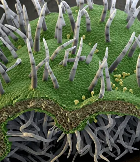
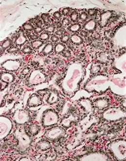
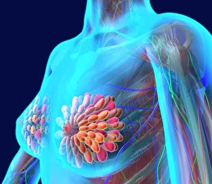
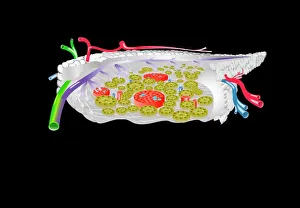
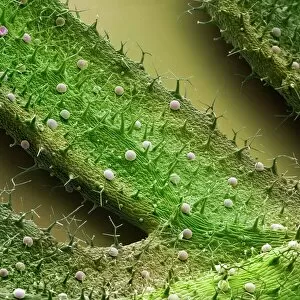
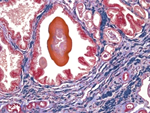
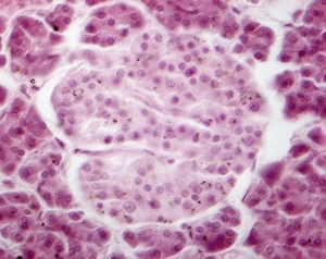
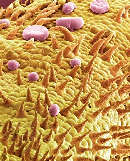
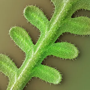
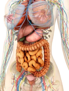
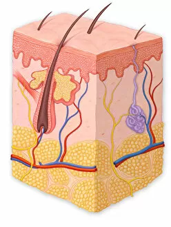
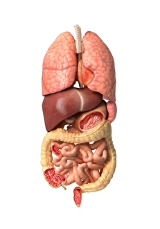
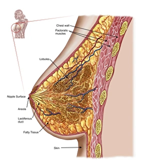
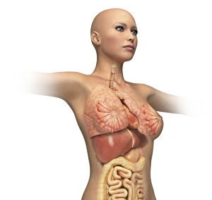
"Gland: The Hidden Marvels of Nature and Anatomy" 1) Botanik Digitalis purpurea L. Fingerhut 160: A captivating glimpse into the intricate beauty of a gland found in the enchanting purple foxglove flower. 2) Lactating breast tissue, light micrograph: Witness the remarkable process of milk production within lactating breast tissue, showcasing the incredible capabilities of mammary glands. 3) Breast anatomy, artwork: Delve into an artistic representation that unveils the complex structure and functionality of the breasts, highlighting their role as vital glands in female physiology. 4) Pancreas anatomy, artwork: Explore a stunning illustration that unravels the inner workings of this crucial gland responsible for regulating blood sugar levels and aiding digestion. 5) French lavender leaf surface, SEM: Uncover the mesmerizing details on a microscopic level as scanning electron microscopy reveals oil glands dotting the surface of a French lavender leaf. 6) Islet of Langerhans, light micrograph: Journey deep into pancreatic tissue to witness these specialized cell clusters known as islets which play a pivotal role in maintaining proper blood glucose levels. 7) Thyroid gland capillaries, SEM: Peer through an electron microscope to marvel at intricately woven networks of tiny blood vessels nourishing one's thyroid gland - an essential player in metabolism regulation. 8) Thyroid gland blood vessels, SEM: Another breathtaking view under high magnification showcases how delicate yet robust blood vessels intertwine within this butterfly-shaped endocrine powerhouse located at our throat's base. 9) Leaf oil glands, SEM: Step inside nature's aromatic treasure troves as scanning electron microscopy captures exquisite images revealing oil glands nestled within leaves - nature's own perfume factories. 10) French lavender leaf, SEM: Get up close and personal with every nook and cranny on a French lavender leaf's surface through scanning electron microscopy - uncovering hidden secrets of this botanical wonder. 11) Kidney anatomy, artwork.