Fraud Blocker Skip to main content

Home > Science > SEM

Climacostomum protozoan, SEM C016 / 9121

 : Climacostomum protozoan, SEM C016  /  9121
In Stock
We accept all major payment options so you can pay for the things you need in the way that is most convenient for you

Choose Your Product

Framed Photos
Buy from $79.99
Photo Prints
Buy from $9.99
Jigsaw Puzzles
Buy from $39.99
Poster Prints
Buy from $24.99
Canvas Prints
Buy from $94.99
Fine Art Prints
Buy from $60.90
Metal Prints
Buy from $119.99
Home Decor
Buy from $16.99

Science Photo Library Photo Prints and Wall Art

Climacostomum protozoan, SEM C016 / 9121

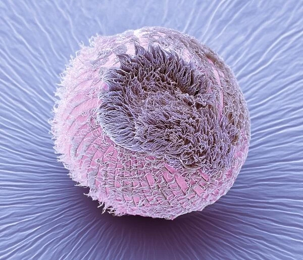
Climacostomum protozoan. Coloured scanning electron micrograph (SEM) of a Climacostomum sp. unicellular ciliate protozoan, showing the cells large oral apparatus (round, centre). This structure features an adoral zone of membranelles (AZM) partly encircling a wide buccal cavity (mouth), which opens into the cytopharyngeal pouch. Magnification: x400 when printed 10 centimetres wide. Science Photo Library features Science and Medical images including photos and illustrations. © STEVE GSCHMEISSNER/SCIENCE PHOTO LIBRARY

Media ID 9245377

Adoral Zone Buccal Cavity Cilia Ciliate Ciliated Cilium Colored Cytopharyngeal Pouch Membranelle Membranelles Micro Organism Micro Organisms Microbiology Microorganism Microorganisms Mouth Oral Apparatus Organism Protist Protozoa Protozoan Protozoans Sphere Spherical Unicellular Microbiological Scanning Electron Microscope

FEATURES IN THESE COLLECTIONS

> Animals > Aquatic > Related Images

> Europe > United Kingdom > Scotland > Highlands > Alness

> Science > SEM

SECURE PAYMENTS
We accept all major payment options so you can pay for the things you need in the way that is most convenient for you

Made in USA
Safe Shipping, Easy Returns

FREE PERSONALISATION
We are proud to offer a range of customisation including Cropping, Captions, and Filters. Zoom Cropping can be adjusted in the basket


EDITORS COMMENTS

This print showcases the intricate beauty of a Climacostomum protozoan, a unicellular ciliate protozoan found in aquatic environments. The coloured scanning electron micrograph (SEM) reveals the remarkable structure of this tiny organism's large oral apparatus at its center. The oral apparatus is composed of an adoral zone of membranelles (AZM), which partially encircles a wide buccal cavity or mouth. This unique feature opens into the cytopharyngeal pouch, allowing for feeding and digestion processes within the cell. At a magnification of x400 when printed 10 centimeters wide, every detail becomes visible, offering us a glimpse into the fascinating world of microbiology. The spherical shape and vibrant colors add to its allure, making it an intriguing subject for scientific study and appreciation. Through this image, we are reminded of the vast diversity that exists within our natural world. It serves as a testament to the complexity and wonder found even in microscopic organisms like Climacostomum protozoans. Such discoveries contribute to our understanding of biology and zoology while highlighting the interconnectedness between all living beings on Earth. Credit: STEVE GSCHMEISSNER/SCIENCE PHOTO LIBRARY.


Framed Prints of Climacostomum protozoan, SEM C016 / 9121

Discover the fascinating world of microorganisms with our Framed Prints from Media Storehouse. This captivating piece features a Coloured Scanning Electron Micrograph (SEM) of a Climacostomum sp. protozoan, captured by Steve Gschmeissner of Science Photo Library. Witness the intricate details of this unicellular ciliate's large oral apparatus, adding an extraordinary touch to your home or office decor. Bring the wonders of science into your living space with our high-quality Framed Prints.


Photo Prints of Climacostomum protozoan, SEM C016 / 9121

Discover the intricacy of the natural world with Media Storehouse's Photographic Prints. Feast your eyes on this captivating Coloured Scanning Electron Micrograph (SEM) of a Climacostomum sp. protozoan, expertly captured by Steve Gschmeissner of Science Photo Library. Witness the mesmerizing detail of this unicellular ciliate's large oral apparatus, a testament to the beauty and complexity of life at the microscopic level. Bring this stunning image into your home or office as a conversation starter and a reminder of the wonders that lie hidden in the smallest corners of our world.


Poster Prints of Climacostomum protozoan, SEM C016 / 9121

Discover the intricacies of the natural world with our Media Storehouse Poster Prints. Featuring the captivating image "Climacostomum protozoan, SEM C016 / 9121" by STEVE GSCHMEISSNER/SCIENCE PHOTO LIBRARY, this poster showcases the colorful detail of a unicellular ciliate protozoan, Climacostomum sp. The large oral apparatus of this fascinating organism takes center stage in this coloured scanning electron micrograph (SEM), revealing its complex structure and the beauty of life at the microscopic level. Bring this mesmerizing image into your home or office to inspire curiosity and ignite conversation.


Jigsaw Puzzles of Climacostomum protozoan, SEM C016 / 9121

Discover the intricacies of the natural world with Media Storehouse's Jigsaw Puzzles. Our latest addition to the collection is this captivating puzzle featuring the Coloured Scanning Electron Micrograph (SEM) of Climacostomum protozoan by STEVE GSCHMEISSNER/SCIENCE PHOTO LIBRARY. Delve into the microscopic realm and assemble the pieces to reveal the stunning details of this unicellular ciliate protozoan's large oral apparatus. A perfect blend of science and fun, this puzzle is an excellent addition to any home or classroom.

diamond
Made With High Grade Materials
thumb_up
Professional Quality Finish
package_2
Carefully Packed To Aid Safe Arrival

You May Also Like


Medieval sunsetMedieval sunset
Add to Basket
EyeUbiquitous_20105148EyeUbiquitous_20105148
Add to Basket
Portrait of a Black-capped chickadee, (Poecile atricapillus) with white backgroundPortrait of a Black-capped chickadee, (Poecile atricapillus) with white background
Add to Basket
Roses with a Blue Tit by a Stream, (oil on panel)Roses with a Blue Tit by a Stream, (oil on panel)
Add to Basket
November Effigies, 1877. Creator: John ThomsonNovember Effigies, 1877. Creator: John Thomson
Add to Basket
The Actor Yamashita Kinsaku II as Lady Kikusui (Kikusui Gozen) in the Play Kaeribana... c. 1779. Creator: Katsukawa ShunkoThe Actor Yamashita Kinsaku II as Lady Kikusui (Kikusui Gozen) in the Play Kaeribana... c. 1779. Creator: Katsukawa Shunko
Add to Basket
Traditional facacde painting on farmhouse in Obertilliach in Lesachtal, East Tyrol, AustriaTraditional facacde painting on farmhouse in Obertilliach in Lesachtal, East Tyrol, Austria
Add to Basket
Lone Tree in WinterLone Tree in Winter
Add to Basket
Lincoln LN3 4 MapLincoln LN3 4 Map
Add to Basket
Palace of Minos, detailed view, Knossos, Heraklion Region, Crete, GreecePalace of Minos, detailed view, Knossos, Heraklion Region, Crete, Greece
Add to Basket
Trades Practiced in Bologna, c1640. Creators: Agostino Parisini, Francesco CurtiTrades Practiced in Bologna, c1640. Creators: Agostino Parisini, Francesco Curti
Add to Basket
Christ on the Mount of Olives, 1428-1429. Creator: Angelico, Fra Giovanni, da Fiesole (around 1400-1455)Christ on the Mount of Olives, 1428-1429. Creator: Angelico, Fra Giovanni, da Fiesole (around 1400-1455)
Add to Basket

Gallery Wall Inspiration

Nerve cell, SEM
Red-barbed ant, SEM
Neutrophil engulfing MRSA, SEM C018 / 8596
Diatom alga, SEM
Diatom, SEM
X and Y chromosomes
Lavender pollen grain, SEM
Scanning Electron Micrograph (SEM): Tardigrade or ‘Water Bear Magnification x 1250 (A4 size: 29. 7 cm width)
Cat tongue surface, SEM
False-coloured SEM (Scanning Electron Micrograph) of a Tardigrade (Tardigrada) amongst moss, from Peak District National Park, Derbyshire, UK. This tardigrade is approximately 0. 1 millimetres long. Digital composite
Collagen, Scanning electron micrograph (SEM)
Santos-Dumont / Sem
T lymphocytes and cancer cell, SEM C001 / 1679
Tardigrade or Water Bear
Hazel pollen grains
Crysotile asbestos
Uterus lining during menstruation, SEM
Iris of the eye, SEM
Stylus in LP record
Salmonella bacteria, SEM
SEM of penicillin fungus
Vascular bundle, SEM
Geranium anther, SEM
Dahlia flower pollen, SEM
Scanning Electron Micrograph (SEM): Praying Mantis - Magnification x 30 (if print A4 size: 29. 7 cm wide)
Tardigrade, SEM
Scanning electron micrograph (SEM) of red blood cell

Discover Our Collections


SEMSEM
View Collection
Aquatic Related ImagesAquatic Related Images
View Collection
AlnessAlness
View Collection


© Copyright Media Storehouse, All Rights Reserved 2003 - 2025
Facebook     Pinterest     100% Payment Secure     Reviews IO