
Protist Collection
"Exploring the Intricate World of Protists: From Seaweed Specimens to Malarial Parasites" In this captivating collection, we delve into the fascinating realm of protists


All Professionally Made to Order for Quick Shipping
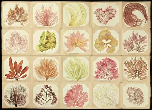
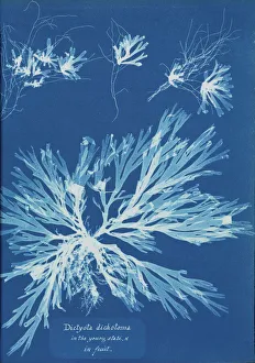
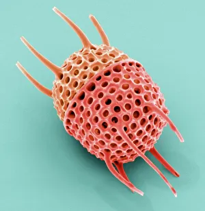
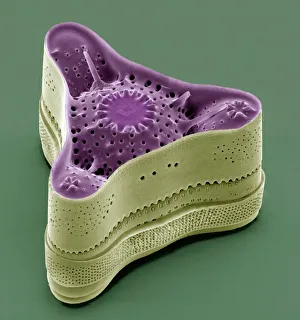
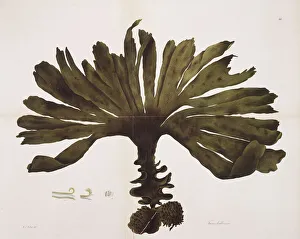
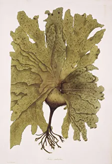
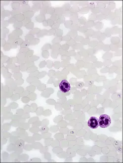
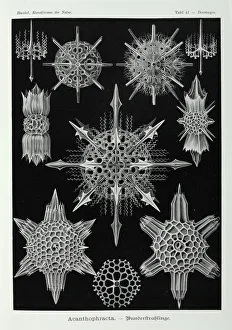
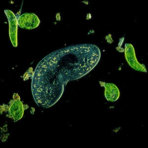
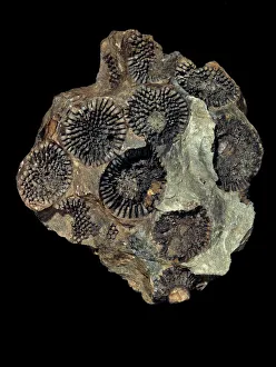
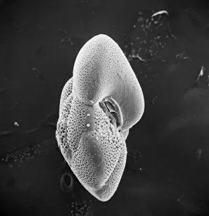
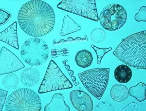
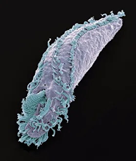
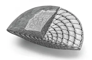
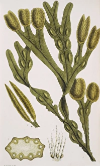
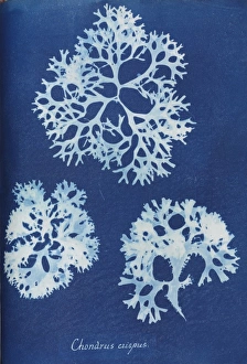
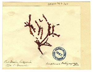
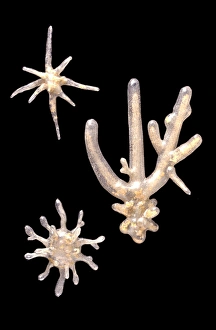
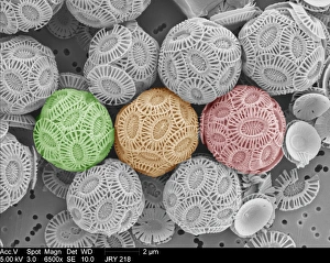
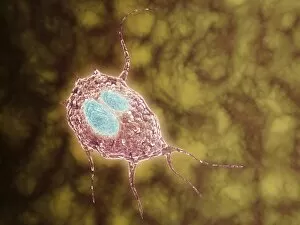
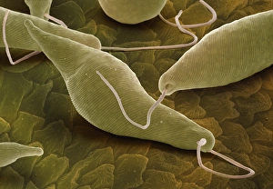
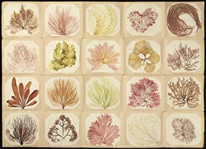
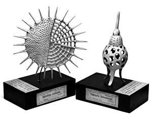
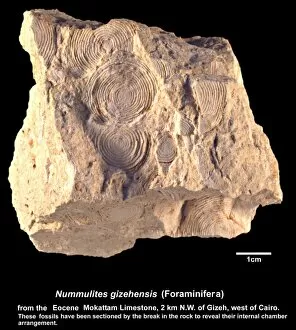
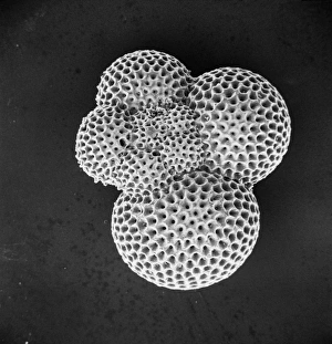
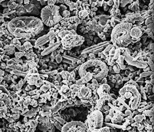
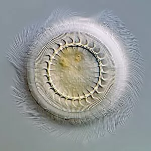
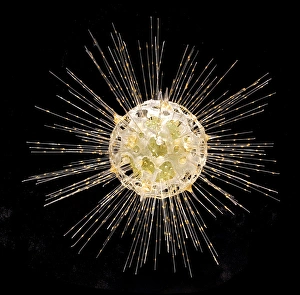
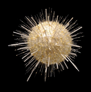
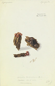
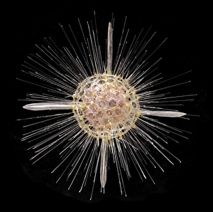
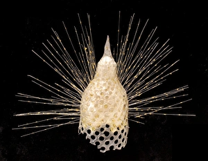
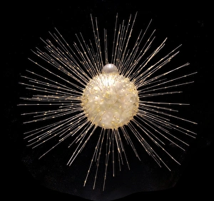
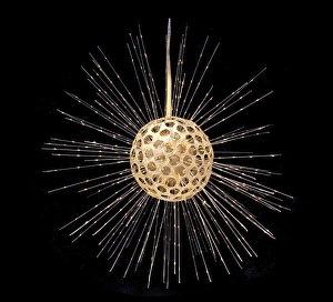
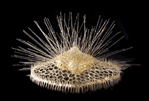
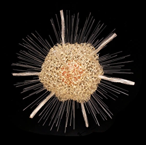
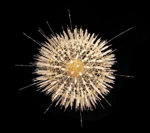
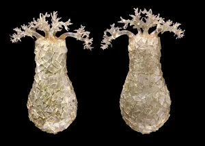
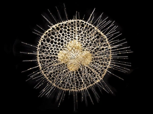
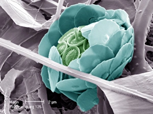
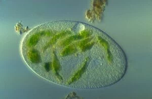
"Exploring the Intricate World of Protists: From Seaweed Specimens to Malarial Parasites" In this captivating collection, we delve into the fascinating realm of protists. The pressed seaweed specimens C016/6127 reveal a stunning diversity, with diatoms taking center stage under the scanning electron microscope (SEM). Dictyota dichotoma showcases its intricate branching structure, reminiscent of an artistic masterpiece. Transporting us back in time is the Rye Beach, New Hampshire Postcard from 1903. This vintage gem captures the beauty of Fucus bulbosus, a majestic kelp swaying in the ocean currents. Another diatom steals our attention under SEM – Fucus radiatus displays its delicate fronds and intricate patterns. But not all they are as visually pleasing; Plasmodium sp. , a malarial parasite, reminds us of their impact on human health. Acanthophracta radiolarians mesmerize us with their intricately sculpted skeletons that resemble miniature works of art. The light micrograph (LM) reveals a hidden world within protozoans - a kidney-shaped ciliate surrounded by Euglena sp. , both magnified x900 when printed A4 size. It's astonishing how much detail can be captured at such high magnification. Calcareous alga Coelosphaeridium adds another dimension to this diverse group while diatoms continue to amaze with their varied shapes and structures. Protists truly showcase nature's creativity and complexity on microscopic scales. Join us as we unravel their secrets and appreciate these tiny wonders that play vital roles in our ecosystems.