Fraud Blocker Skip to main content

Microorganism Collection

Microorganisms, the tiny life forms that exist all around us, are truly fascinating

Background imageMicroorganism Collection: HeLa cells, light micrograph C017 / 8299

HeLa cells, light micrograph C017 / 8299
HeLa cells, multiphoton fluorescence micrograph (MFM). The cell nuclei, which contain the cells genetic information, are purple. Microtubules are blue and actin microfilaments are red

Background imageMicroorganism Collection: Neutrophil engulfing MRSA, SEM C018 / 8596

Neutrophil engulfing MRSA, SEM C018 / 8596
Neutrophil engulfing MRSA. Coloured scanning electron micrograph (SEM) of a neutrophil white blood cell (green) engulfing methicillin-resistant Staphylococcus aureus bacteria (MRSA, pink)

Background imageMicroorganism Collection: HeLa cells, light micrograph C017 / 8298

HeLa cells, light micrograph C017 / 8298
HeLa cells, multiphoton fluorescence micrograph (MFM). The cell nuclei, which contain the cells genetic information, are blue. Golgi bodies, which modify and package proteins, are orange

Background imageMicroorganism Collection: Water bear, light micrograph C016 / 8581

Water bear, light micrograph C016 / 8581
Water bear, differential interference contrast (DIC) light micrograph. Water bears, or tardigrades, (phylum Tardigrada) are small, water-dwelling

Background imageMicroorganism Collection: Microscopic view of human respiratory syncytial virus

Microscopic view of human respiratory syncytial virus (RSV). RSV causes respiratory tract infection of the lungs and breathing passages

Background imageMicroorganism Collection: Infections spread by sneezing, artwork C013 / 5949

Infections spread by sneezing, artwork C013 / 5949
Infections spread by sneezing. Computer artwork showing virus particles (virions, blue) and bacteria (rod-shaped) in the spray produced by someone when they sneeze

Background imageMicroorganism Collection: Microscopic view of chlamydia

Microscopic view of chlamydia. Chlamydia is a common sexually transmitted disease (STD) caused by a bacterium

Background imageMicroorganism Collection: Flu virus particle, artwork F008 / 3245

Flu virus particle, artwork F008 / 3245
Flu virus particle, computer artwork. In the virus envelope are two types of protein spike, haemagglutinin (H, pink) and neuraminidase (N, orange), which determine the strain of virus

Background imageMicroorganism Collection: Plankton, artwork C016 / 3447

Plankton, artwork C016 / 3447
Plankton. Artwork of plankton as viewed under a magnifying glass

Background imageMicroorganism Collection: Salmonella bacteria, artwork C013 / 8818

Salmonella bacteria, artwork C013 / 8818
Salmonella bacteria, computer artwork. Salmonella sp. bacteria are gram-negative rod-shaped bacteria that have flagella (hair-like structures) that they use for locomotion

Background imageMicroorganism Collection: Water bear, SEM C016 / 9084

Water bear, SEM C016 / 9084
Water bear (Echiniscus granulatus). Coloured scanning electron micrograph (SEM) showing the underside of a water bear, or tardigrade (phylum Tardigrada)

Background imageMicroorganism Collection: Bloom

Bloom
Takumi Takemura

Background imageMicroorganism Collection: In Bloom

In Bloom
Takumi Takemura

Background imageMicroorganism Collection: Bloom

Bloom
Takumi Takemura

Background imageMicroorganism Collection: Colony of Haemophilus influenzae, 1906 (litho)

Colony of Haemophilus influenzae, 1906 (litho)
3501914 Colony of Haemophilus influenzae, 1906 (litho) by German School, (20th century); Private Collection; (add.info.: Bacteria constitute a large domain of prokaryotic microorganisms)

Background imageMicroorganism Collection: Colony of Mycobacterium leprae, 1906 (litho)

Colony of Mycobacterium leprae, 1906 (litho)
3501912 Colony of Mycobacterium leprae, 1906 (litho) by German School, (20th century); Private Collection; (add.info.: Bacteria constitute a large domain of prokaryotic microorganisms)

Background imageMicroorganism Collection: Colony of Micrococcus Gonorrhoea, 1906 (litho)

Colony of Micrococcus Gonorrhoea, 1906 (litho)
3501903 Colony of Micrococcus Gonorrhoea, 1906 (litho) by German School, (20th century); Private Collection; (add.info.: Bacteria constitute a large domain of prokaryotic microorganisms)

Background imageMicroorganism Collection: Colony of Streptococcus pneumoniae with and without bubble capsule, 1906 (litho)

Colony of Streptococcus pneumoniae with and without bubble capsule, 1906 (litho)
3501899 Colony of Streptococcus pneumoniae with and without bubble capsule, 1906 (litho) by German School, (20th century); Private Collection; (add.info)

Background imageMicroorganism Collection: Colony of Streptococcus Pneumoniae, 1906 (litho)

Colony of Streptococcus Pneumoniae, 1906 (litho)
3501898 Colony of Streptococcus Pneumoniae, 1906 (litho) by German School, (20th century); Private Collection; (add.info.: Bacteria constitute a large domain of prokaryotic microorganisms)

Background imageMicroorganism Collection: Spirillum of the Lyme disease causing bacteria, Spirochaetes Borrelia Recurrentis

Spirillum of the Lyme disease causing bacteria, Spirochaetes Borrelia Recurrentis, in blood sample, 1906 (litho)
3501919 Spirillum of the Lyme disease causing bacteria, Spirochaetes Borrelia Recurrentis, in blood sample, 1906 (litho) by German School

Background imageMicroorganism Collection: Colony of Vibrio cholerae, 1906 (litho)

Colony of Vibrio cholerae, 1906 (litho)
3501918 Colony of Vibrio cholerae, 1906 (litho) by German School, (20th century); Private Collection; (add.info.: Bacteria constitute a large domain of prokaryotic microorganisms)

Background imageMicroorganism Collection: Colony of Clostridium tetani with spores, 1906 (litho)

Colony of Clostridium tetani with spores, 1906 (litho)
3501913 Colony of Clostridium tetani with spores, 1906 (litho) by German School, (20th century); Private Collection; (add.info.: Bacteria constitute a large domain of prokaryotic microorganisms)

Background imageMicroorganism Collection: Colony of Mycobacterium tuberculosis, 1906 (litho)

Colony of Mycobacterium tuberculosis, 1906 (litho)
3501910 Colony of Mycobacterium tuberculosis, 1906 (litho) by German School, (20th century); Private Collection; (add.info.: Bacteria constitute a large domain of prokaryotic microorganisms)

Background imageMicroorganism Collection: A colony of Staphylococcus pyogenes after cell division, 1906 (litho)

A colony of Staphylococcus pyogenes after cell division, 1906 (litho)
3501897 A colony of Staphylococcus pyogenes after cell division, 1906 (litho) by German School, (20th century); Private Collection; (add.info)

Background imageMicroorganism Collection: Colony of Streptococcus pyogenes, 1906 (litho)

Colony of Streptococcus pyogenes, 1906 (litho)
3501894 Colony of Streptococcus pyogenes, 1906 (litho) by German School, (20th century); Private Collection; (add.info.: Bacteria constitute a large domain of prokaryotic microorganisms)

Background imageMicroorganism Collection: Varieties of microscopic Marine organisms, 1900 (litho)

Varieties of microscopic Marine organisms, 1900 (litho)
3501931 Varieties of microscopic Marine organisms, 1900 (litho) by German School, (20th century); Private Collection; Prismatic Pictures

Background imageMicroorganism Collection: Colony of Yersinia pestis, 1906 (litho)

Colony of Yersinia pestis, 1906 (litho)
3501917 Colony of Yersinia pestis, 1906 (litho) by German School, (20th century); Private Collection; (add.info.: Bacteria constitute a large domain of prokaryotic microorganisms)

Background imageMicroorganism Collection: Colony of Salmonella Typhi showing Bacilli with flagella, 1906 (litho)

Colony of Salmonella Typhi showing Bacilli with flagella, 1906 (litho)
3501916 Colony of Salmonella Typhi showing Bacilli with flagella, 1906 (litho) by German School, (20th century); Private Collection; (add.info)

Background imageMicroorganism Collection: Colony of Salmonella typhi, 1906 (litho)

Colony of Salmonella typhi, 1906 (litho)
3501915 Colony of Salmonella typhi, 1906 (litho) by German School, (20th century); Private Collection; (add.info.: Bacteria constitute a large domain of prokaryotic microorganisms)

Background imageMicroorganism Collection: Colony of Bacillus Anthracis in a human spleen, 1906 (litho)

Colony of Bacillus Anthracis in a human spleen, 1906 (litho)
3501905 Colony of Bacillus Anthracis in a human spleen, 1906 (litho) by German School, (20th century); Private Collection; (add.info)

Background imageMicroorganism Collection: Spores of Bacillus Anthracis, 1906 (litho)

Spores of Bacillus Anthracis, 1906 (litho)
3501904 Spores of Bacillus Anthracis, 1906 (litho) by German School, (20th century); Private Collection; (add.info.: Bacteria constitute a large domain of prokaryotic microorganisms)

Background imageMicroorganism Collection: Colony of Micrococcus Gonorrhoea found in the Urethra, 1906 (litho)

Colony of Micrococcus Gonorrhoea found in the Urethra, 1906 (litho)
3501900 Colony of Micrococcus Gonorrhoea found in the Urethra, 1906 (litho) by German School, (20th century); Private Collection; (add.info)

Background imageMicroorganism Collection: Colony of Corynebacterium Diphtheriae, 1906 (litho)

Colony of Corynebacterium Diphtheriae, 1906 (litho)
3501907 Colony of Corynebacterium Diphtheriae, 1906 (litho) by German School, (20th century); Private Collection; (add.info.: Bacteria constitute a large domain of prokaryotic microorganisms)

Background imageMicroorganism Collection: Colony of Bacterium mallei, 1906 (litho)

Colony of Bacterium mallei, 1906 (litho)
3501906 Colony of Bacterium mallei, 1906 (litho) by German School, (20th century); Private Collection; (add.info.: Bacteria constitute a large domain of prokaryotic microorganisms)

Background imageMicroorganism Collection: Staphylococcus colony, 1905 (litho)

Staphylococcus colony, 1905 (litho)
3501895 Staphylococcus colony, 1905 (litho) by German School, (20th century); Private Collection; (add.info.: Bacteria constitute a large domain of prokaryotic microorganisms)

Background imageMicroorganism Collection: Colony of Mycobacterium Tuberculosis, 1906 (litho)

Colony of Mycobacterium Tuberculosis, 1906 (litho)
3501911 Colony of Mycobacterium Tuberculosis, 1906 (litho) by German School, (20th century); Private Collection; (add.info.: Bacteria constitute a large domain of prokaryotic microorganisms)

Background imageMicroorganism Collection: The liar

The liar
Coloured scanning electron micrograph (SEM) of a deer ked (Lipoptena cervi). L. cervi lives as an ectoparasite on deer, moose and other cervids

Background imageMicroorganism Collection: Alien wearing a face mask, illustration

Alien wearing a face mask, illustration

Background imageMicroorganism Collection: Histopathology and pathophysiology of diabetic food ulcers

Histopathology and pathophysiology of diabetic food ulcers

Background imageMicroorganism Collection: Scanning electron micrograph of HIV particles infecting a human H9 T cell

Scanning electron micrograph of HIV particles infecting a human H9 T cell

Background imageMicroorganism Collection: Coxiella burnetii, the bacteria that causes Q Fever

Coxiella burnetii, the bacteria that causes Q Fever. A dry fracture of a Vero cell exposing the contents of a vacuole where Coxiella burnetii are busy growing

Background imageMicroorganism Collection: Stylized rabies virus particles

Stylized rabies virus particles, the cause of the viral neuroinvasive disease acute encephalitis

Background imageMicroorganism Collection: Microscopic view of Neisseria gonorrhoeae

Microscopic view of Neisseria gonorrhoeae. Neisseria gonorrhoeae typically infects the mucous membranes causing infections such as urethritis, cervicitis, salpingitis, pelvic inflammatory disease

Background imageMicroorganism Collection: Microscopic view of yellow fever virus

Microscopic view of yellow fever virus. Yellow fever is an acute viral disease

Background imageMicroorganism Collection: Microscopic view of Giardiasis

Microscopic view of Giardiasis, an infectious disease caused by a unicellular parasite known as Giardia lamblia

Background imageMicroorganism Collection: Microscopic view of bacterial pneumonia

Microscopic view of bacterial pneumonia. Bacterial pneumonia is a type of pneumonia caused by bacterial infection. Pneumonia can be generally defined as inflammation of the lung parenchyma

Background imageMicroorganism Collection: Microscopic view of Streptococcus pneumoniae

Microscopic view of Streptococcus pneumoniae, also known as pneumococcus. Pneumococcus is a Gram-positive coccus shaped pathogenic bacteria which causes many types of pneumococcal infections in

Background imageMicroorganism Collection: Anatomy of foot fungus with microscopic close-up

Anatomy of foot fungus with microscopic close-up


All Professionally Made to Order for Quick Shipping

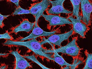
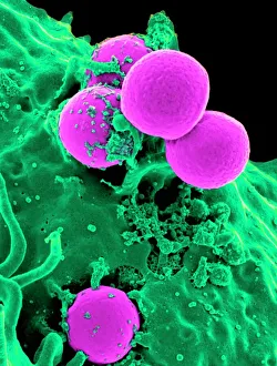
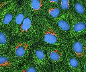
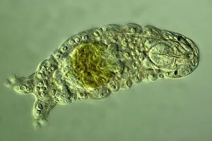
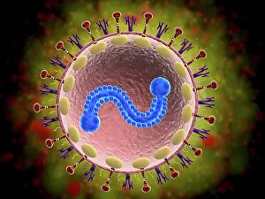
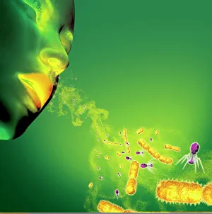
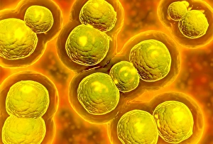
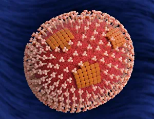
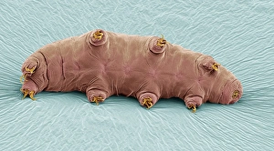
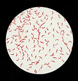
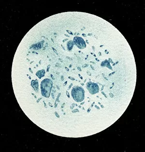
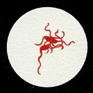
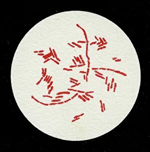
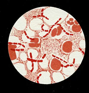
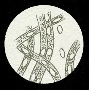
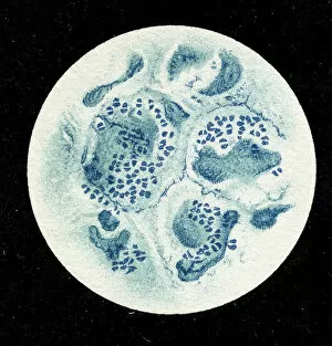
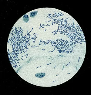
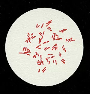
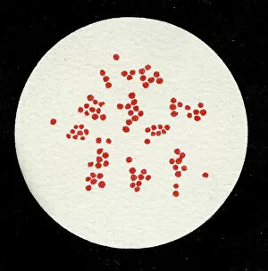
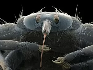
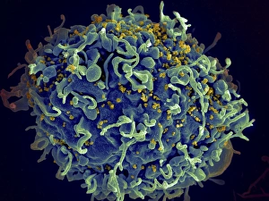
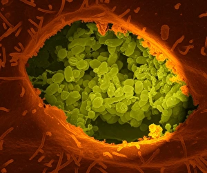
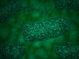
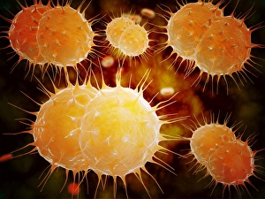
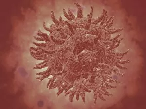
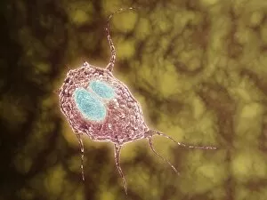
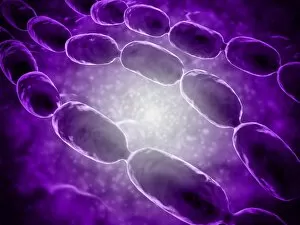
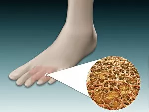
Microorganisms, the tiny life forms that exist all around us, are truly fascinating. From HeLa cells to water bears and viruses like MRSA and RSV, these microscopic entities play a significant role in our lives. In the world of science, HeLa cells have been instrumental in numerous breakthroughs. Captured under a light micrograph (C017 / 8299), their intricate structures reveal the complexity of human biology. Another captivating image shows neutrophils engulfing MRSA bacteria, as seen through a scanning electron microscope (C018 / 8596). This powerful defense mechanism showcases how our immune system fights against harmful invaders. Water bears, also known as tardigrades, are remarkable creatures that can survive extreme conditions. A light micrograph (C016 / 8581) captures their unique appearance with their stout bodies and claw-like limbs. Equally intriguing is another image taken using a scanning electron microscope (SEM C016 / 9084), which reveals even more details about these resilient organisms. Viruses such as respiratory syncytial virus and flu virus particles have caused widespread infections throughout history. While one can only be observed under a microscope (microscopic view of human respiratory syncytial virus), the other is depicted through artwork showcasing its structure (flu virus particle, artwork F008 / 3245). These images remind us of the importance of vaccinations in preventing viral outbreaks. The spread of infections by sneezing is illustrated through an artistic representation (artwork C013 / 5949). It serves as a reminder to practice good hygiene to protect ourselves and others from contagious diseases. Chlamydia may be small but has significant consequences for those affected by it. A microscopic view highlights its presence within host cells—a stark reminder of the need for awareness and prevention efforts against sexually transmitted infections. Looking back at historical lithographs from 1906 brings attention to colonies formed by Haemophilus influenzae and Mycobacterium leprae.