
Protozoans Collection
Protozoans are fascinating microscopic organisms that inhabit various environments, from freshwater to soil


All Professionally Made to Order for Quick Shipping
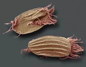
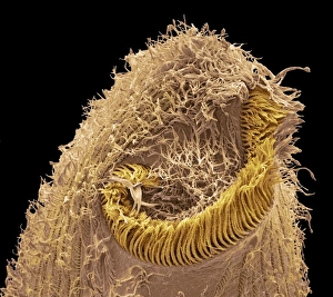
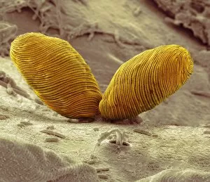
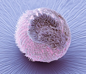
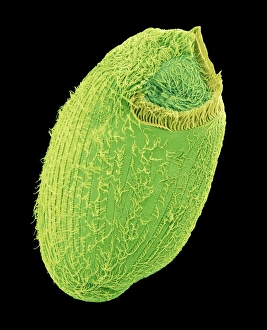
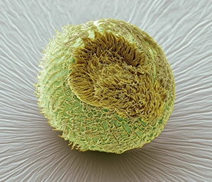
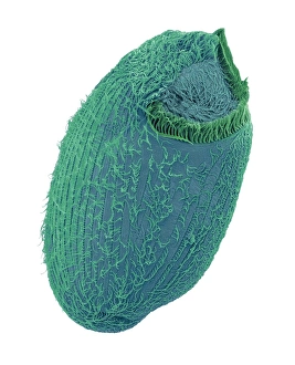
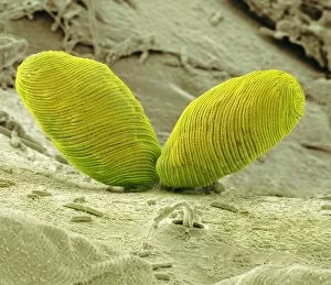
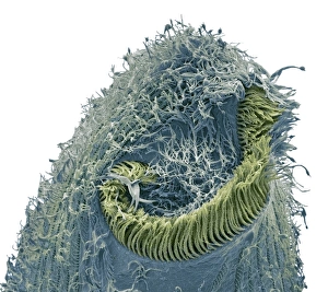
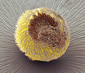
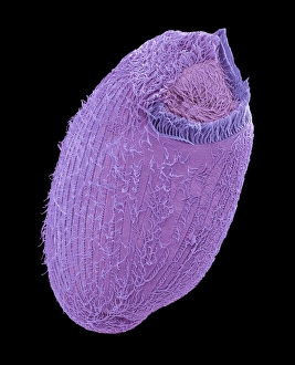
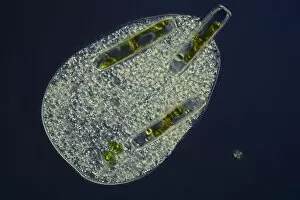
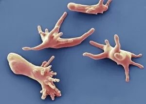
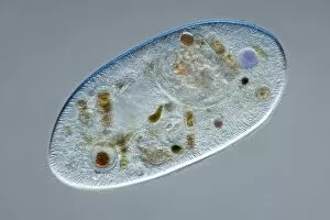
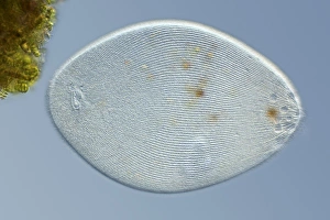
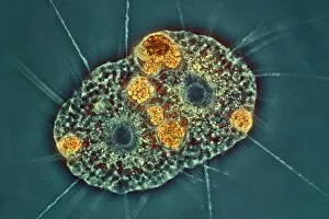
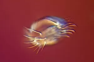
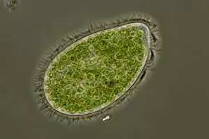
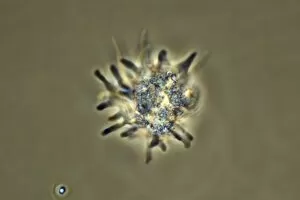
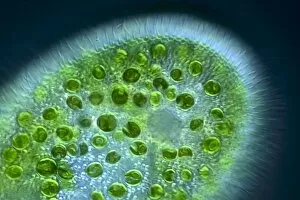
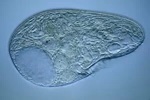
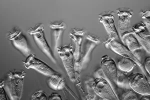
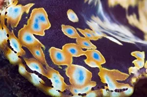
Protozoans are fascinating microscopic organisms that inhabit various environments, from freshwater to soil. These incredible creatures come in a wide array of shapes and sizes, each with its own unique characteristics. One captivating example is the Holosticha ciliate protozoan, as seen under a scanning electron microscope (SEM). Its intricate body structure and delicate cilia make it a mesmerizing sight to behold. Similarly, the Euplotes protozoa, also observed through SEM C016 / 9040 imaging, showcases its remarkable complexity. Another intriguing species is the Climacostomum protozoan. SEM images such as C016 / 9063 and C016 / 9121 reveal its distinctive features and provide insight into its behavior within its environment. The Climacostomum's ability to adapt and survive in diverse conditions is truly awe-inspiring. The Hartmannella vermiformis protozoa cysts captured in image C016 / 9401 demonstrate their resilient nature. These cysts serve as protective structures during unfavorable conditions until they can resume their active life cycle. Euglena flagellate protozoa are another group worth mentioning due to their fascinating flagella-driven locomotion. SEM images like C016 / 9103 and C016 / 9104 showcase these graceful organisms moving through their aquatic habitats with ease.