Premium Framed Print : Human head, MRI and 3D CT scans C018 / 0636


![]()
Framed Photos From Science Photo Library
Human head, MRI and 3D CT scans C018 / 0636
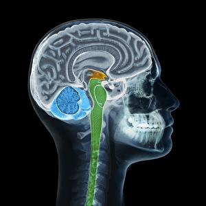
Human head. Coloured composite image of a magnetic resonance imaging (MRI) scan of the brain and 2D and 3D computed tomography (CT) scans of the head and neck of a 35 year old patient. The highly folded area of the brain is the cerebrum, which is responsible for conscious thought, emotion and voluntary movement. The light grey arch beneath it is the corpus callosum, which facilitates communication between the hemispheres. At bottom right is the cerebellum, which controls muscle coordination and balance
Science Photo Library features Science and Medical images including photos and illustrations
Media ID 9236835
© ZEPHYR/SCIENCE PHOTO LIBRARY
Bones Cerebellum Cervical Spine Composite Ct Scan Diagnosis Diagnostic Facial Mri Scan Neck Profile Radiography Saggital Scanner Thirties Thirty Five Three Dimensional Vertebra X Ray Machine Xray Brain Section Sectioned
14"x16" Premium Frame
Contemporary style Premium Wooden Frame with 8"x10" Print. Complete with 2" White Mat and 1.25" thick MDF frame. Printed on 260 gsm premium paper. Glazed with shatter proof UV coated acrylic glass. Backing is paper covered backing with rubber bumpers. Supplied ready to hang with a pre-installed sawtooth/wire hanger. Care Instructions: Spot clean with a damp cloth. Securely packaged in a clear plastic bag and envelope in a reinforced cardboard shipper
FSC Real Wood Frame and Double Mounted with White Conservation Mountboard - Professionally Made and Ready to Hang
Estimated Image Size (if not cropped) is 25.4cm x 20.3cm (10" x 8")
Estimated Product Size is 40.6cm x 35.6cm (16" x 14")
These are individually made so all sizes are approximate
Artwork printed orientated as per the preview above, with landscape (horizontal) orientation to match the source image.
EDITORS COMMENTS
This print showcases the intricate details of a human head, combining magnetic resonance imaging (MRI) and computed tomography (CT) scans. The composite image features a 35-year-old patient's brain, neck, and facial profile in stunning color against a black background. The highly folded area visible in the brain scan is known as the cerebrum, responsible for conscious thought, emotion, and voluntary movement. Just beneath it lies the light grey arch called the corpus callosum, which plays a crucial role in facilitating communication between the brain's hemispheres. At the bottom right corner of the image rests the cerebellum – an essential component controlling muscle coordination and balance. This visual masterpiece provides valuable insights into human anatomy and serves as a powerful tool for medical diagnosis. It highlights various sections of organs such as bones within both head and neck regions while offering an anatomical perspective on normal bodily structures. With its three-dimensional representation achieved through advanced radiography techniques like MRI scanning and CT imaging using x-ray machines, this print captures not only biological complexity but also showcases cutting-edge technology at work. ZEPHYR/SCIENCE PHOTO LIBRARY has skillfully captured this remarkable fusion of artistry and science to create an awe-inspiring piece that appeals to both medical professionals seeking diagnostic clarity and individuals fascinated by our incredible human body.
MADE IN THE USA
Safe Shipping with 30 Day Money Back Guarantee
FREE PERSONALISATION*
We are proud to offer a range of customisation features including Personalised Captions, Color Filters and Picture Zoom Tools
SECURE PAYMENTS
We happily accept a wide range of payment options so you can pay for the things you need in the way that is most convenient for you
* Options may vary by product and licensing agreement. Zoomed Pictures can be adjusted in the Basket.