
Mri Scan Collection
"Exploring the Intricate Brain Fibres: Unveiling Insights through MRI Scans" MRI scans have revolutionized our understanding of brain anatomy


All Professionally Made to Order for Quick Shipping
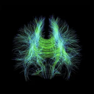
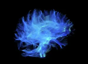
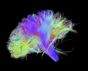
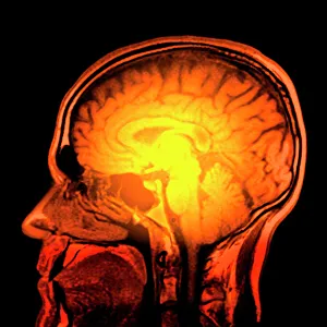
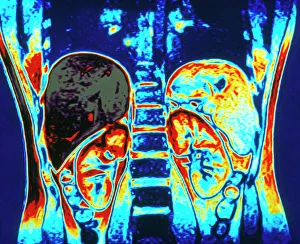
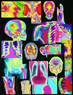
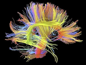
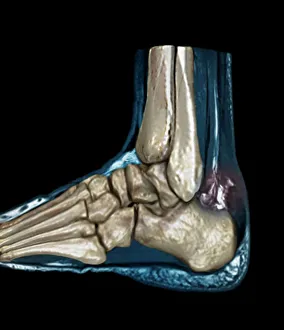
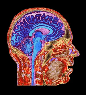
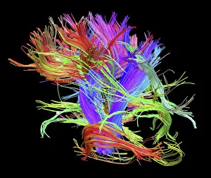
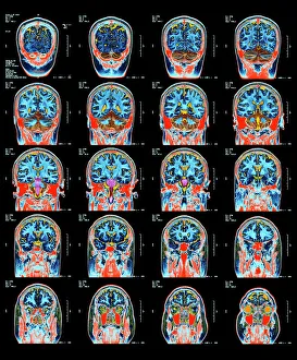
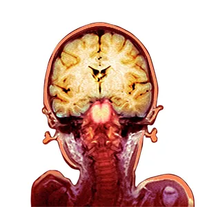
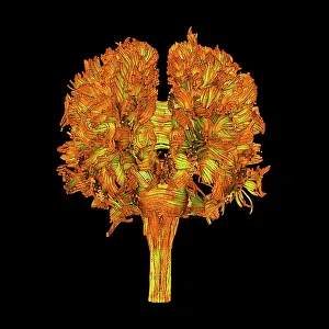
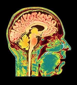
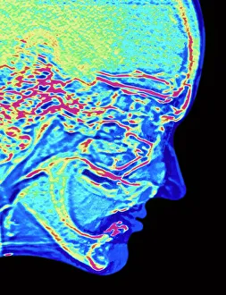
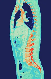
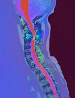
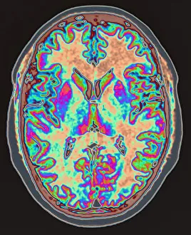
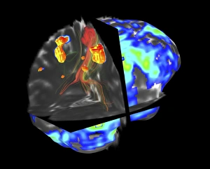
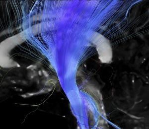
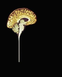
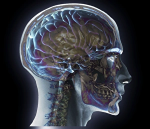
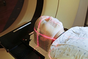
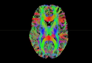
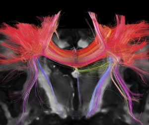
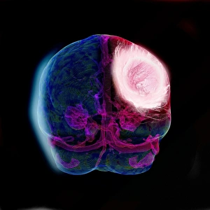
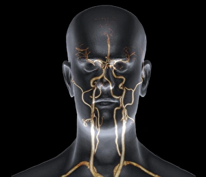
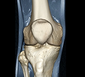
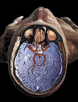
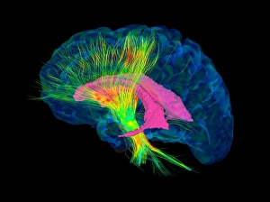
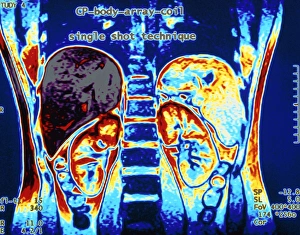
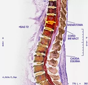
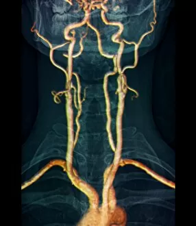
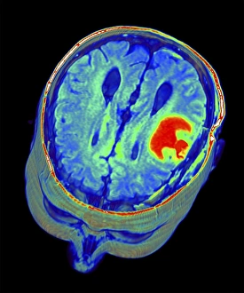
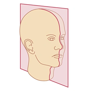
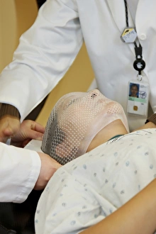
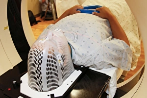
"Exploring the Intricate Brain Fibres: Unveiling Insights through MRI Scans" MRI scans have revolutionized our understanding of brain anatomy, allowing us to delve deep into the intricate web of brain fibres. With advanced techniques like DTI (Diffusion Tensor Imaging), such as in C017 / 7099 and C017 / 7035 scans, we can visualize the complex pathways that connect different regions of the brain. Intriguingly, white matter fibres play a crucial role in transmitting information across these neural networks. The mesmerizing images captured in C014 / 5666 and C014 / 5668 offer a glimpse into this vital aspect of human brain function. But they are not limited to studying brains alone. They enable us to explore other parts of the body too. A vivid example is the colour MRI scan showcasing kidneys and liver within the abdomen – a testament to medical imaging's versatility. Beyond just organs, an assortment of coloured X-rays and body scans provides healthcare professionals with valuable insights for diagnosis and treatment planning. These comprehensive tools empower them to make informed decisions regarding patient care. Sometimes, unfortunate incidents occur that necessitate an MRI scan for accurate assessment. For instance, ruptured Achilles tendon cases like C018 / 0649 or breast implant complications may require detailed examination using MRI technology. On a brighter note, healthy individuals also benefit from routine brain MRIs (C016 / 8845). By establishing baseline images, doctors can monitor any changes over time and ensure optimal neurological well-being. MRI scans continue to be at the forefront of medical imaging advancements – unraveling mysteries hidden beneath our skin with remarkable precision. As we unlock more secrets about our bodies through these cutting-edge technologies, their impact on healthcare becomes increasingly profound.