Fraud Blocker Skip to main content

Scanner Collection

"Unveiling the Hidden Wonders: The Scanner's Journey" Delicate and ethereal

Background imageScanner Collection: Full body scan, MRI scan

Full body scan, MRI scan
Full body scan of a female swimmer. Her brain is purple (top), her heart and arteries are red/orange (upper centre), her kidneys are pale green (centre)

Background imageScanner Collection: Sea green swallowtail butterfly

Sea green swallowtail butterfly
Sea green swallowtail (Papilio lorquinianus) butterfly. This butterfly is found in Indonesia, Sulawesi, Borneo, Java, Sumatra and in the Philippines

Background imageScanner Collection: Brain anatomy, MRI scan

Brain anatomy, MRI scan
Brain anatomy. Coloured magnetic resonance imaging (MRI) scan of the human head from the side. The sagittal scan has halved the brain, revealing the internal anatomy

Background imageScanner Collection: Fingerprint scanner

Fingerprint scanner. Persons finger being scanned by a fingerprint scanner. This scanner is being used to open a door. The ridges on peoples fingers form a unique identifying pattern

Background imageScanner Collection: Ruptured Achilles tendon, MRI C018 / 0649

Ruptured Achilles tendon, MRI C018 / 0649
Ruptured Achilles tendon. Coloured magnetic resonance imaging (MRI) scan of the foot of a 25 year old patient with a ruptured Achilles tendon

Background imageScanner Collection: Fingerprint scanner, artwork

Fingerprint scanner, artwork
Fingerprint scanner, computer artwork

Background imageScanner Collection: Normal human brain, MRI scan C016 / 8845

Normal human brain, MRI scan C016 / 8845
Brain. Coloured magnetic resonance imaging (MRI) scan of a sagittal section through a patients head showing a healthy human brain

Background imageScanner Collection: Ruptured breast implants, MRI

Ruptured breast implants, MRI
Ruptured breast implants. Coloured magnetic resonance imaging (MRI) scan of ruptures in both of a womans breast implants (red). These are intracapsular ruptures, the most common type

Background imageScanner Collection: Childs brain, MRI scan

Childs brain, MRI scan
Childs brain. Coloured magnetic resonance imaging (MRI) scan of an coronal (vertical) section through the head and brain of a healthy 3-year-old child, passing through the ears

Background imageScanner Collection: White matter fibres, DTI scan

White matter fibres, DTI scan
White matter fibres. Coloured 3D diffusion tensor imaging (DTI) scan of the brain, showing the bundles of white matter nerve fibres

Background imageScanner Collection: Normal human brain, MRI scan C016 / 8850

Normal human brain, MRI scan C016 / 8850
Brain. Coloured magnetic resonance imaging (MRI) scan of a sagittal section through a patients head showing a healthy human brain

Background imageScanner Collection: Normal torso, MRI scan

Normal torso, MRI scan
Normal torso, coloured profile (sagittal) magnetic resonance imaging (MRI) scan. The front of the body is at left. The spinal bones (vertebrae)

Background imageScanner Collection: Inflamed spinal discs, MRI scan

Inflamed spinal discs, MRI scan
Inflamed spinal discs. Coloured magnetic resonance imaging (MRI) scan of a side view of the spine of a patient with discitis. The front of the body is at left, the skull is at top

Background imageScanner Collection: Model Gillian Duxbury in the E. M. I. X-Ray scanner with the team from L / R Godfrey

Model Gillian Duxbury in the E. M. I. X-Ray scanner with the team from L / R Godfrey
Model Gillian Duxbury in the E.M.I. X-Ray scanner with the team from L/R Godfrey Hounsfield (The inventor of the scanner), Tony Williams (Mechanical Designer), Peter Langstone (Electronics expert)

Background imageScanner Collection: At an aircraft receiving depot, cadets are being x-rayed in modern manner

At an aircraft receiving depot, cadets are being x-rayed in modern manner, miniature film being used. The films after development are projected on to a screen and cases of suspected T.B

Background imageScanner Collection: Model Gillian Duxbury in E. M. I. X-Ray scanner. April 1975 75-1905-006

Model Gillian Duxbury in E. M. I. X-Ray scanner. April 1975 75-1905-006
Model Gillian Duxbury in E.M.I. X-Ray scanner. April 1975 75-1905-006

Background imageScanner Collection: Baird Electron Scanner System of Television

Baird Electron Scanner System of Television, showing the Electron Scanner when used for the televising of talking films. This can be employed for a definition of 100-500 lines

Background imageScanner Collection: Diane Jackman of Stockton is the first patient on the new scanner at North Tees Hospital

Diane Jackman of Stockton is the first patient on the new scanner at North Tees Hospital, with senior radiographer John Elliott. 30th June 1993

Background imageScanner Collection: Eye-prints, 1938

Eye-prints, 1938. Churchmans cigarette card, from a series titled Modern Wonders [WA & AC Churchman, Great Britain & Ireland, 1938]

Background imageScanner Collection: Stroke, MRI and 3D CT scans C016 / 6419

Stroke, MRI and 3D CT scans C016 / 6419
Stroke. Coloured composite image of a magnetic resonance imaging (MRI) scan of the brain and 2D and 3D computed tomography (CT)

Background imageScanner Collection: Normal human brain, MRI scan C016 / 8843

Normal human brain, MRI scan C016 / 8843
Brain. Coloured magnetic resonance imaging (MRI) scan of a sagittal section through a healthy human brain. The highly folded area (brown) is the cerebrum, which is responsible for conscious thought

Background imageScanner Collection: Normal skull and brain, 3D CT scan C016 / 6333

Normal skull and brain, 3D CT scan C016 / 6333
Normal skull and brain. Coloured 3D computed tomography (CT) scan of the head of a 35 year old patient. Part of the skull has been cut-away to show the brain (purple and red)

Background imageScanner Collection: Normal head and neck, MRI and 3D CT scans C016 / 6337

Normal head and neck, MRI and 3D CT scans C016 / 6337
Normal head. Coloured composite image of a magnetic resonance imaging (MRI) scan of the brain and a 3D computed tomography (CT) scan of the head and neck of a 35 year old patient

Background imageScanner Collection: Human torso, MRI scan

Human torso, MRI scan
Normal human torso, monochrome profile magnetic resonance imaging (MRI) scan. The front of the body is at left. The spinal bones (vertebrae) are seen running from the top of the chest (top)

Background imageScanner Collection: Lasers used to position a patient wearing a short face mask in a computed tomography

Lasers used to position a patient wearing a short face mask in a computed tomography
Lasers are used to position a patient model wearing a short face mask in a computed tomography (CT) scanner to obtain images of a tumor before radiation therapy

Background imageScanner Collection: Neck and head arteries, MRA scan C016 / 6449

Neck and head arteries, MRA scan C016 / 6449
Neck and head arteries. Coloured magnetic resonance angiography (MRA) scan of a normal carotid system of a 36 year old patient

Background imageScanner Collection: Brain cancer, DTI and 3D CT scans C016 / 6414

Brain cancer, DTI and 3D CT scans C016 / 6414
Brain cancer. Coloured composite image of diffusion tensor imaging (DTI) scans of the brains, and 3D computed tomography (CT) scans of the skulls, of two 35 year old patients

Background imageScanner Collection: External carotid artery, 3D CT scan C016 / 6341

External carotid artery, 3D CT scan C016 / 6341
External carotid artery. Coloured 3D computed tomography (CT) angiogram (blood vessel scan) of the head showing a normal external carotid artery (red, in neck) and its branches

Background imageScanner Collection: Uterine fibroids, MRI scan C018 / 0466

Uterine fibroids, MRI scan C018 / 0466
Uterine fibroids. Magnetic resonance imaging (MRI) scan of the pelvis of a 47 year old female with two large fibroids (centre) in her uterus

Background imageScanner Collection: Healthy knee, CT scan C018 / 0413

Healthy knee, CT scan C018 / 0413
Healthy knee. Coloured frontal computed tomography (CT) scan projected on to a magnetic resonance imaging (MRI) scan of the knee of a 30 year old

Background imageScanner Collection: Brain blood supply, MRI scan C018 / 0432

Brain blood supply, MRI scan C018 / 0432
Brain haemorrhage. Coloured magnetic resonance imaging (MRI) scan of the brain of a 49 year old patient with a subdural haematoma (red)

Background imageScanner Collection: Artery damage, MRA scan

Artery damage, MRA scan
Artery damage. Magnetic resonance angiography (MRA) scan of the neck of a 23-year-old male motorcycle crash victim. Dissection of the right internal carotid artery (left)

Background imageScanner Collection: Polycystic kidney, ultrasound scan C017 / 7746

Polycystic kidney, ultrasound scan C017 / 7746
Polycystic kidney. Ultrasound scan of the kidney of a 42 year old female showing numerous fluid-filled cysts (dark areas)

Background imageScanner Collection: Aortic dissection, 3D CT scan

Aortic dissection, 3D CT scan
Aortic dissection. 3D computed tomography (CT) scan of a 67-year-old man with an aortic dissection. The aorta, the bodys main artery, is running vertically down centre (red)

Background imageScanner Collection: Multiple sclerosis, MRI scan

Multiple sclerosis, MRI scan

Background imageScanner Collection: Brain tumour, 3D-MRI scan

Brain tumour, 3D-MRI scan
Brain tumour. Coloured three-dimensional model of a male head, made up from numerous magnetic resonance imaging (MRI) scans

Background imageScanner Collection: COMPUTER ROOM, 1999. Disc storage systems at the Cape Cod Air Station in Massachusetts

COMPUTER ROOM, 1999. Disc storage systems at the Cape Cod Air Station in Massachusetts. Photograph by Bob Kramer, 1999

Background imageScanner Collection: beauty, biometric, boot, checking, color image, computer graphic, concept, copy space

beauty, biometric, boot, checking, color image, computer graphic, concept, copy space
Woman using biometric scanners in futuristic corridor

Background imageScanner Collection: Radiation therapists fit a short face mask to a patient during radiation therapy

Radiation therapists fit a short face mask to a patient during radiation therapy
Two radiation therapists fit a short face mask to a patient model to steady the head during radiation therapy

Background imageScanner Collection: Patient positioned in computed tomography scanner

Patient positioned in computed tomography scanner
A patient model wearing a short face mask is positioned in a computed tomography (CT) scanner to obtain images of a tumor before radiation therapy

Background imageScanner Collection: Domestic Dog, mongrel, adult, being scanned for microchip by vet at veterinary surgery, England

Domestic Dog, mongrel, adult, being scanned for microchip by vet at veterinary surgery, England, February

Background imageScanner Collection: Multispectral scanner reveals lithologic and structural features of Great Namaland

Multispectral scanner reveals lithologic and structural features of Great Namaland in Namibia with clarity. Area extremely dry with little vegetation, so geology dominates image

Background imageScanner Collection: Sheep farming, ultrasound scanner screen, scanning ewes during early stage pregnancy to see how

Sheep farming, ultrasound scanner screen, scanning ewes during early stage pregnancy to see how many lambs they are carrying, England, November

Background imageScanner Collection: Polycystic kidneys, ultrasound scan

Polycystic kidneys, ultrasound scan
Polycystic kidneys. Ultrasound scan of the kidney of an 82 year old male showing numerous fluid-filled cysts (dark areas)

Background imageScanner Collection: Brain scan, MRI scan F006 / 4629

Brain scan, MRI scan F006 / 4629
Normal brain. Coloured magnetic resonance imaging (MRI) scan of an axial section through a healthy brain. The front of the brain is towards top of the image

Background imageScanner Collection: Brain scan, MRI scan F006 / 4610

Brain scan, MRI scan F006 / 4610
Normal brain. Coloured magnetic resonance imaging (MRI) scan of an axial section through a healthy brain. The front of the brain is towards top of the image

Background imageScanner Collection: Brain scan, MRI scan F006 / 4617

Brain scan, MRI scan F006 / 4617
Normal brain. Coloured magnetic resonance imaging (MRI) scan of an axial section through a healthy brain. The front of the brain is towards top of the image

Background imageScanner Collection: Brain scan, MRI scan F006 / 4607

Brain scan, MRI scan F006 / 4607
Normal brain. Coloured magnetic resonance imaging (MRI) scan of an axial section through a healthy brain. The front of the brain is towards top of the image


All Professionally Made to Order for Quick Shipping

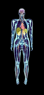
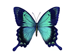
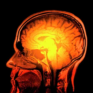
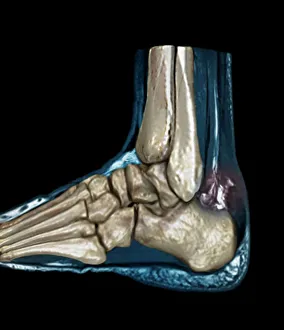
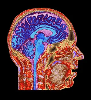
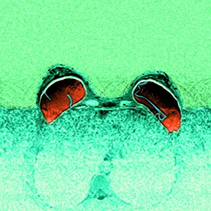
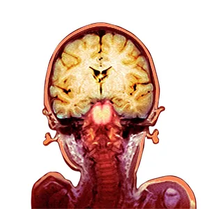
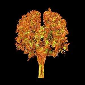
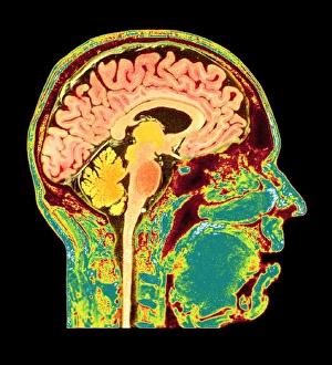
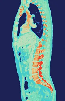
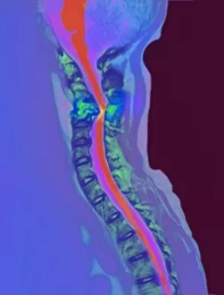
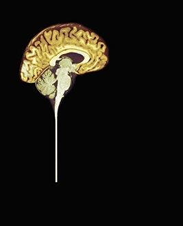
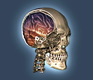
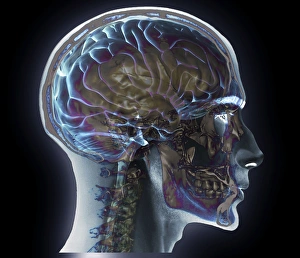
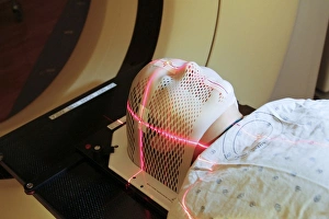
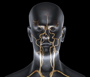
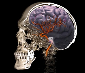
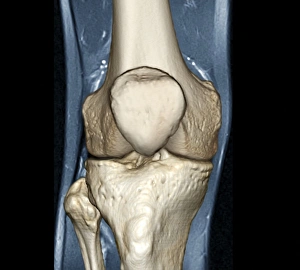
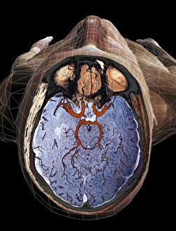
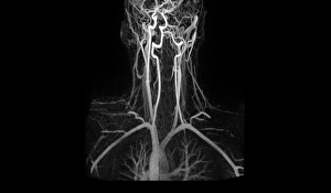
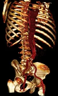
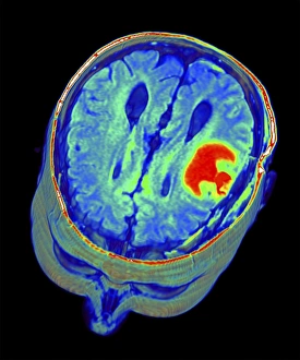
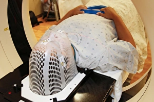
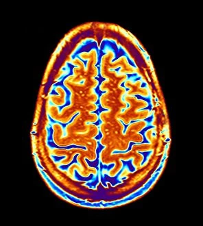
"Unveiling the Hidden Wonders: The Scanner's Journey" Delicate and ethereal, a sea green swallowtail butterfly gracefully flutters its wings under the watchful eyes of a scanner. With precision and accuracy, a full body scan captures every detail, revealing the intricate workings of our complex human form. Peering into the depths of brain anatomy, an MRI scan unveils the mysteries hidden within, unlocking secrets that shape our thoughts and actions. Like a unique signature, a fingerprint scanner maps out individual patterns with meticulous care, ensuring security in an ever-evolving world. Injured but not defeated, an MRI C018 / 0649 reveals the extent of damage to a ruptured Achilles tendon - guiding doctors towards effective treatment options. Beyond fingerprints' utilitarian purpose lies artistic beauty; an artwork created by capturing each swirl through a fingerprint scanner tells tales untold before. A glimpse into normalcy is found as an MRI C016 / 8845 showcases the intricacies of a healthy human brain - reminding us of its remarkable resilience. Seeking answers beneath layers unseen, an MRI exposes ruptured breast implants - providing clarity for those seeking resolution and healing. Innocence encapsulated within imaging technology as we witness childlike wonder through glimpses into young minds via captivating MRI scans. Unraveling connections that make us who we are; white matter fibers come alive under DTI scans - illuminating pathways crucial for communication within our brains. Echoing familiarity amidst diversity; another snapshot from everyday life emerges with Normal Human Brain captured by MRI C016 / 8850 – reminding us how extraordinary ordinary can be.