Pillow > Arts > Street art graffiti > Digital art > Digital paintings
Pillow : Histopathology and pathophysiology of diabetic food ulcers


![]()
Home Decor From Stocktrek
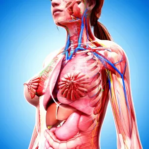
Histopathology and pathophysiology of diabetic food ulcers
Stocktrek Images specializes in Astronomy, Dinosaurs, Medical, Military Forces, Ocean Life, & Sci-Fi
Media ID 13002313
© Enid Hajderi/Stocktrek Images
Abnormal Anatomy Arteriole Artery Biology Biomedical Illustrations Blood Blood Cells Blood Flow Blood Vessels Bloodstream Capillaries Cardiovascular System Cell Cell Biology Chart Circulation Circulatory System Cross Section Cutaway View Cytology Dermis Disease Erythrocyte Feet Haematid Healthcare Hematology Hemoglobin Human Foot Human Tissue Human Toes Illness Infection Leukocyte Macrophage Magnification Medical Medicine Microbiology Microorganism Microscopic Molecular Biology Oxygen Pathology Physiology Problem Red Blood Cells Red Blood Corpuscles Red Cells Text Tube Western Script Wound Angiogenesis Diabetes Fibroblast Healing Histopathology Lesion Necrosis Periosteum Ulcer
18"x18" (46x46cm) Pillow
18"x18" (46x46cm) Faux Suede Pillow with a plush soft feel. Your choice of image fills the front, with a stone colored faux suede back. Flat sewn concealed white zip.
Accessorise your space with decorative, soft pillows
Estimated Product Size is 45.7cm x 45.7cm (18" x 18")
These are individually made so all sizes are approximate
Artwork printed orientated as per the preview above, with landscape (horizontal) or portrait (vertical) orientation to match the source image.
FEATURES IN THESE COLLECTIONS
> Arts
> Street art graffiti
> Digital art
> Digital paintings
> Maps and Charts
> Related Images
> Posters
> Aircraft Posters
> Cutaway Posters
EDITORS COMMENTS
This print titled "Histopathology and Pathophysiology of Diabetic Foot Ulcers" offers a visually stunning depiction of the intricate biomedical illustrations related to this medical condition. The color image, presented in a horizontal format, showcases a digitally generated cutaway view of the human foot and toes. The artwork delves into the histopathology and pathophysiology underlying diabetic foot ulcers, providing valuable insights into the physiology and pathology involved. The cross section reveals various components such as red blood cells, blood vessels, capillaries, arterioles, leukocytes, macrophages, microorganisms, fibroblasts, and more. Through meticulous magnification and close-up views within this illustration's composition, viewers can observe the detailed structures like periosteum (bone covering), dermis (skin layer), subcutis (fat layer), as well as cellular elements including erythrocytes (red blood cells) with their hemoglobin content. This scientifically accurate representation sheds light on how diabetes affects wound healing processes by impacting circulation and oxygen supply to cells. It emphasizes the importance of understanding molecular biology within our circulatory system for effective healthcare management. Enid Hajderi's artwork serves as an invaluable resource for researchers in fields such as medicine microbiology or cytology. Its educational value lies in its ability to elucidate complex concepts surrounding diabetic foot ulcers while showcasing the beauty found within biological systems.
MADE IN THE USA
Safe Shipping with 30 Day Money Back Guarantee
FREE PERSONALISATION*
We are proud to offer a range of customisation features including Personalised Captions, Color Filters and Picture Zoom Tools
SECURE PAYMENTS
We happily accept a wide range of payment options so you can pay for the things you need in the way that is most convenient for you
* Options may vary by product and licensing agreement. Zoomed Pictures can be adjusted in the Basket.