
Artery Collection
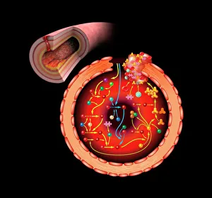
"Exploring the intricate pathways of life: The artery, a vital conduit for our body's functions


All Professionally Made to Order for Quick Shipping
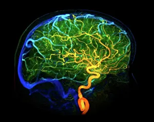
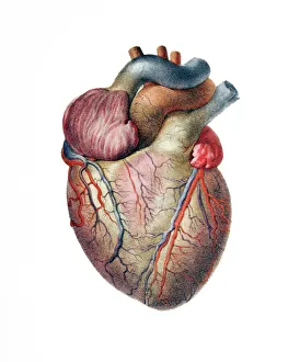
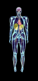
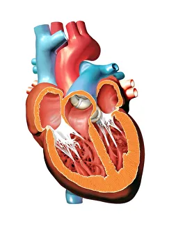
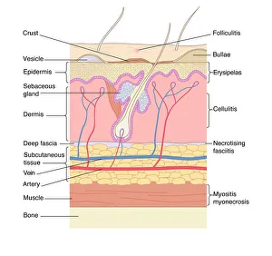
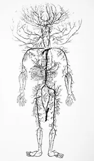
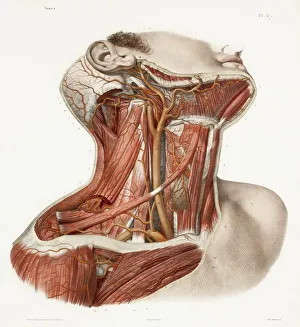
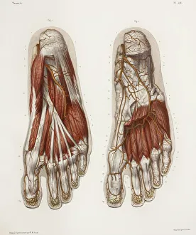
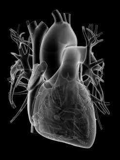
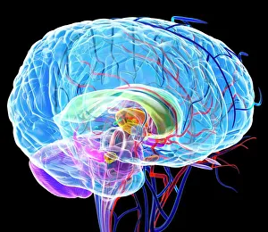
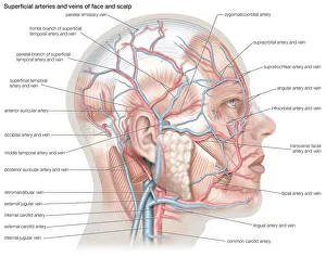
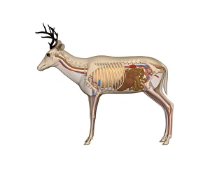
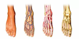
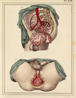
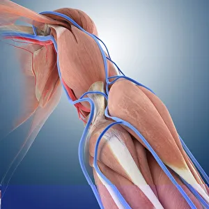
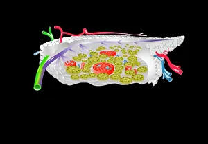
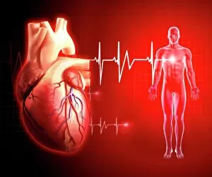
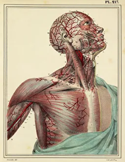
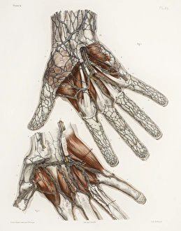
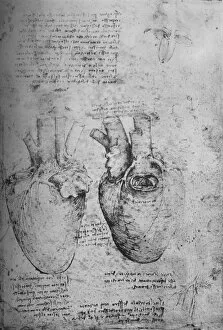
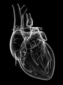
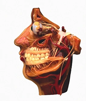
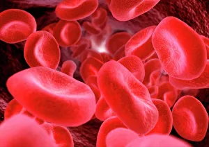
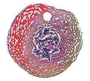
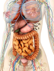
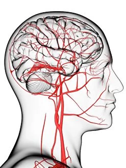
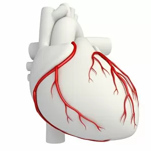
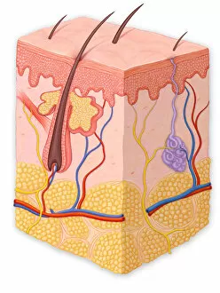
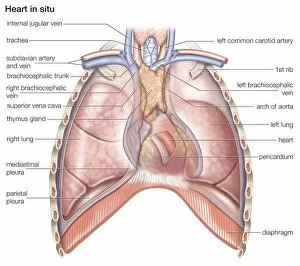
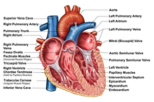
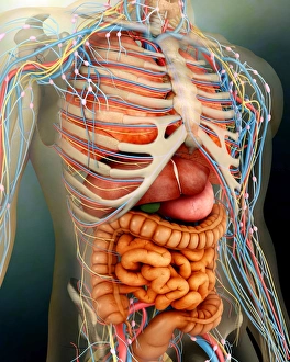
"Exploring the intricate pathways of life: The artery, a vital conduit for our body's functions. " Delving into the depths of our brain's blood vessels, we uncover the mesmerizing intricacies that keep us thinking and dreaming. Witnessing the wonders of medical technology through a 3D angiogram C007/1981, we marvel at how science unravels the mysteries within our arteries. The heart, an emblem of love and vitality, relies on its network of arteries to pump life-giving blood throughout our entire being. Peering beneath the surface with a full-body scan or MRI scan reveals a symphony of arteries orchestrating health and well-being in every corner. Human heart anatomy comes alive through breathtaking artwork, reminding us of this organ's crucial role in sustaining life itself. Exploring skin disorders through artistic depictions serves as a reminder that even our outermost layer is intimately connected to arterial health. Tracing back centuries ago, an 18th-century illustration showcases early insights into understanding the complex arterial system that sustains us all. Historical artwork depicting neck vascular anatomy takes us on a journey through time while appreciating how far medical knowledge has come today. Our brain's blood vessels hold secrets waiting to be discovered - their delicate structure supporting cognition and consciousness alike. Gazing deep into retinal veins and arteries reminds us that even our eyes rely on these tiny highways for clear vision and visual perception. X-rays reveal neck and shoulder arteries like hidden rivers flowing silently beneath our skin - nurturing muscles and bones with oxygen-rich blood. Stepping back in time with a 19th-century illustration showcasing foot anatomy highlights how even seemingly distant extremities depend on healthy arterial circulation. 13. Artistic renderings unveil the captivating complexity of brain anatomy – where countless neurons thrive thanks to nourishment from dedicated arterial pathways.